Combination 830-nm and 633-nm light-emitting diode phototherapy shows promise in the treatment of recalcitrant psoriasis: preliminary findings

Photomedicine and Laser Surgery Volume 00, Number 00, 2009ª Mary Ann Liebert, Inc.
Pp. 1–6DOI: 10.1089=pho.2009.2484 Combination 830-nm and 633-nm Light-Emitting Diode Phototherapy Shows Promise in the Treatment of Recalcitrant Psoriasis: Preliminary Findings Glynis Ablon, M.D., FAAD Background and Objectives: Psoriasis is one of the major problems facing dermatologists worldwide. Planararrays of light-emitting diodes (LEDs) have recently attracted attention in the treatment of difficult dermato-logical entities, 830 nm in near infrared (near-IR) and 633 nm in visible red. This study was designed to assess theefficacy of combination 830-nm and 633-nm LED phototherapy in the treatment of recalcitrant psoriasis. Subjectsand Methods: Nine informed and consenting patients with psoriasis were enrolled in this preliminary study,(3 men, 6 women, mean age 34.3, skin types I to IV). All had chronic psoriasis, which in most cases had provedresistant to conventional treatments. They were treated sequentially with LED arrays delivering continuous-wave 830 nm (near-IR) and 633 nm (red) in two 20-min sessions over 4 or 5 weeks, with 48 h between sessions(830 nm, 60 J=cm2; 633 nm, 126 J=cm2). Results: All patients completed their LED regimens (4 requiring 1 regi-men, 5 requiring a second). Follow-up periods were from 3 to 8 months, except in two patients who were lost tofollow-up. Clearance rates at the end of the follow-up period ranged from 60% to 100%. Satisfaction wasuniversally very high. Conclusions: The antiinflammatory effects of LED energy at 830 nm and 633 nm have beenwell documented, as has their use in wound healing. LED phototherapy is easy to apply, pain free and side-effect free, and is well tolerated by patients of all skin types. The promising results of this preliminary studywarrant a proper controlled double-blind study with a larger patient population.
some streptococcal infections, and activation of the epidermaldendritic Langerhans cell, an antigen-presenting cell, by an One of the major problem diseases facing dermatolo- as-yet-unknownantigen,isbelievedtoplayamajorroleinthe gists in clinical practice is psoriasis, affecting between psoriatic process.
2% and 3% of the population worldwide, despite advances in Phototherapy has played a historical role in the treatment the understanding of its etiology and development of new of psoriasis; exposure to sunlight, in small quantities to treatment approaches. Psoriasis (from the Greek psora mean- prevent sunburn, is known to improve psoriasis. The pho- ing ‘to itch') has been recognized from biblical times and was tosensitizer psoralen plus ultraviolet A light (PUVA) was even at one time confused with leprosy (Greek lepra, scaly popular a decade and more ago and is being used even now, skin, þ psora). Although there are several phenotypes, plaque although long-term follow-up has elicited extremely unde- psoriasis or psoriasis vulgaris accounts for approximately sirable side effects such as the appearance of cutaneous 90% of cases. Psoriasis was originally believed to be a disorder melanomas.3 The excimer laser has also been used with some primarily associated with epidermal keratinocytes leading to success, although the areas treated are small, so large pla- the typical inflammation and itchy hyperkeratotic plaques, ques take a long time to treat, and the same potential exists but recent research has added an immunological component for UV-related carcinogenic side effects. Various pharma- to the equation, with CD4 þ T-cells that have turned ‘‘rogue,'' cotherapeutic topical and oral systemic treatments have been attacking instead of defending normal skin.1,2 Various trig- used, but the potential for side effects with long-term use, gers have been identified, including stress, skin injury, and such as steroid-mediated skin atrophy and liver damage, is Ablon Skin Institute, Los Angeles, California.
high. However, if treated incorrectly or left untreated, the demographics, history of psoriasis, and details of previous appearance of psoriatic comorbidities has been noted, such treatment regimens are seen in Table 1. All patients, having as loss of quality of life leading to onset of depressive illness, had the purpose of the study explained to them, signed psoriatic arthritis, and even cardiovascular disease.4 consent forms to participate in the study and for clinical Phototherapy with non-UV light sources might prove inter- photography. The Ethics Committee of the Ablon Skin In- esting. The wavelength of 633 nm in the visible red waveband stitute approved the study, which was conducted under the has been associated with good results in the treatment of acne precepts of the Declaration of Helsinki (Rev 5, 2000). None of vulgaris, another disease with a T-cell-mediated immune com- the subjects had any history of reactivity to visible or near-IR ponent,5,6 and 830 nm in the near-infrared (near-IR) waveband light, and at the time of the study, none was using any kind has strong antiinflammatory effects;7,8 830 nm has also been of potentially photosensitizing medication.
shown to induce the production of peripheral endogenous opi-oids,9 thereby potentially relieving itching. A new generation of System and treatment regimen planar-mounted light-emitting diodes (LEDs) has attracted at-tention in a wide spectrum of specialities, and one such system The system used was the Omnilux from Photo Ther- offers arrays delivering continuous light at 830 nm and 633 nm.
apeutics (Carlsbad, CA) fitted with the plus and revive The present preliminary study was designed to assess the effect heads, delivering 830 nm (near-IR) and 633 nm (visible red), of the combination of 830 nm and 633 nm LED phototherapy, respectively, in a continuous wave. The head used was at- applied sequentially, in the treatment of cases of chronic psori- tached to the base, and the articulated panels that make up asis that were recalcitrant to conventional therapies.
the head were arranged to follow as closely as possible thecontour of the area being treated and positioned approxi- Subjects and Methods mately 5 cm from the skin. Each single LED session lasted20 min, giving incident radiant fluences of 60 J=cm2 for the 830-nm head and 126 J=cm2 for the 633-nm head. The stan- The subjects in the study included nine psoriasis patients dard treatment regimen lasted for 5 weeks with 10 treatment who attended the author's institute for the treatment of their sessions made up as follows: near-IR head on the first day psoriasis. Eight cases had plaque psoriasis, and there was followed 48 h later by another near-IR treatment in week 1; one case of guttate psoriasis. There were three men and six visible red head on the first day followed 48 h later by an- women, with a mean age of 34.3  11.2 (range 22–58) and other visible red treatment in week 2; and near-IR treatment skin types from I to IV. All had psoriasis (body surface area followed by visible red treatment 48 h later each week in involvement range 5–80%, 4 months to 35 years duration) weeks, 3, 4, and 5. If 100% resolution was achieved before the that had proved resistant to conventional treatments. Patient full 5-week regimen, LED phototherapy was stopped. If Table 1. Patient Demographics, Psoriasis History, and Prior Treatment Psoriasis history 35 years, 60% BSA (back PUVA, methotrexate, acitretin, and torso, upper and alefacept, topical treatments lower extremities, and (steroids, vitamin D, tazarotene gel.
groin), recent onset Moderate improvement of psoriatic arthritis (50% BSA) but withunacceptable side effects 14 years, 80% BSA Methotrexate (failed), topical treatments (poor response) Recent onset, 30% BSA Topical treatments, calcipotriene cream(minimal effect) 1 years, 15% BSA (scalp, elbows, Olux, tazarotene gel knees, posterior neck) 3 years, 5% BSA (elbows, knees) Ultraviolet B (no tar, failed), currently taking fluocinonide(with steroid atrophy around plaques) 5 years, 50% BSA, guttate psoriasis Cephalexin, calcipotriene and betamethasone dipropionate(3 weeks), clearance except shins 4 months, 60% BSA (back, buttocks, Topical treatments, but no result elbows, knees & scalp) 1 year, 15 BSA (back, elbows) Calcipotriene and betamethasone dipropionate for 5 months,minimal improvement 6 years, 40% BSA (elbows, knees, Topical fluocinonide, PUVA, shins, buttocks, back) BSA, body surface area; PUVA, psoralen plus ultraviolet A phototherapy.
LED PHOTOTHERAPY FOR PSORIASIS patients were already participating in a topical regimen and the topical was not a photosensitizer, in most cases thedosage was reduced during LED phototherapy. If clearance All nine patients completed their respective treatment was insufficient, or if the patient requested it, a second reg- protocols. No adverse side effects were noted, and patients imen was indicated, with the same treatment protocol as the felt no pain during the treatment. Five required two first regimen. Patients were asked to return for regular fol- regimens, and the remaining four needed only one regimen.
low-up sessions up to a maximum of 8 months. For typical Two patients (Patients 1 & 2) required the full 5-week, 10- home-applied adjunctive care, patients were recommended treatment regimen; the others had 4 weeks with eight treat- to use nonprescription over-the-counter hydrophilic oint- ments. Resolution rates ranged from 60% to 100% (mean ments or oils daily to keep the plaques moist.
91.7  13.7%), and follow-up periods ranged from 3 to 8 Table 2. Details of Light-Emitting Diode (LED) Treatments Per Session, Per Regimen, and Results with Follow-Up LED treatment per single regimen* (regimens required, n) Clearance and post-treatment follow-up Wk 1: 2830 nm; Wk 2: 2633 nm; Wk 3, 4, 5: 1st regimen: 50% (thighs), 90% (back), 80% recurrence 1830 nm & 1633 nm (2) seen 11 months post-treatment 2nd regimen: 60% (thighs), 100% (back), no recurrence in 3 months þ topical treatments Test treatment on large plaque right anterior shin 100% clearance, maintained with calcipotriene and Wk 1: 2830 nm; Wk 2: 2633 nm; betamethasone dipropionate & tazarotene gel for Wk 3, 4, 5: 1830 nm 1633 nm (1) 8 months. No further LED treatment required Test treatment on largest plaques on elbows & knees 1st regimen: 80% clearance, maintained without Wk 1: 2830 nm; Wk 2: 2633 nm; any other treatment for 2 months, when Wk 3, 4: 1830 nm & 1633 nm (2) 2nd regimen: 100% clearance, but patient lost to follow-up after 6 weeks because moved out of state Clobetasol propionate, tazarotene gel started 100% clearance by 4th treatment week. Completely concomitantly with LED treatment clear 4 months post-treatment Wk 1: 2830 nm; Wk 2: 2633 nm;Wk 3, 4: 1830 nm & 1633 nm (1) Wk 1: 2830 nm; Wk 2: 2633 nm; Wk 3, 4: 1830 nm 1st regimen: 50% clearance with resveratrol-containing emollient cream plus tazarotene gel. Patient wanted2nd LED treatment 2nd regimen: 100% clearance 2 weeks post-treatment, tazarotene gel discontinued during LED treatment.
Completely clear at 6-month follow-up Wk 1: 2830 nm; Wk 2: 2633 nm; 1st regimen: 75% clearance, patient requested further Wk 3, 4: 1830 nm & 1633 nm (2) 2nd regimen: 3 sessions only, 1830 nm, 1633 nm, 1830 nm, 48-h rest between each. No topicals usedwith LED treatment. 90% clearance of treatedlesions, maintained < 10% BSA at 6 monthspost-treatment with no form of medication Recommended to have LED treatment by friend; 1st regimen: plus calcipotriene and betamethasone only back and buttocks treated with LED dipropionate and tazarotene gel, 50% clearance Wk 1: 2830 nm; Wk 2: 2633 nm; Patient requested further LED treatment regimen Wk 3, 4: 1830 nm & 1633 nm (2) 2nd regimen: 90% clearance after 3rd week; 95% resolution after final week on back andbuttocks, maintained at 80% 6 months after finaltreatment with topicals. Other areas treated onlywith topical treatments remain at 40% improvement Wk 1: 2830 nm; Wk 2: 2633 nm; Reduced calcipotriene and betamethasone Wk 3, 4: 1830 nm & 1633 nm (1) dipropionate dose, added tazarotene gel. After 2ndweek, 60% clearance, improved to 90% 3 weekspost-treatment. At 3-month follow-up, 95%resolution maintained with topical calcipotrieneand betamethasone dipropionate in a.m. only Wk 1: 2830 nm; Wk 2: 2633 nm; Calcipotriene and betamethasone dipropionate Wk 3, 4: 1830 nm & 1633 nm (1) started with start of LED treatment. 2 weekspost-treatment, 60% resolution. Patient did notreturn; lost to follow-up *Unless otherwise stated, 48-h rest period between weekly LED sessions and second LED regimen same as first regimen.


months, except in two patients who were lost to follow-up at2 (Patient 9) and 6 weeks (Patient 3). Patient 9 (60% clear-ance) failed to turn up for any further follow-up sessions.
Patient 3 (100% resolution) moved to the other side of thecountry but was extremely satisfied with the results. In allpatients with the longer follow-up periods, satisfaction rateswere universally high. Details of treatment regimens, clear-ance rates, pharmacotherapy, and follow-up periods areshown in Table 2.
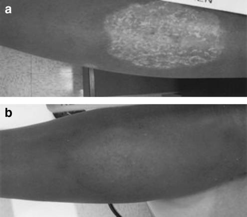
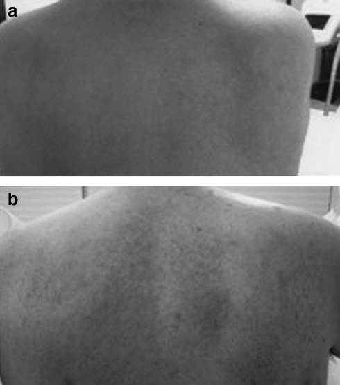
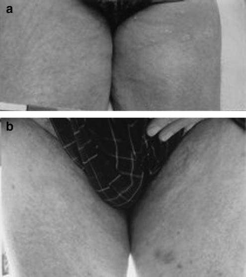
Figures 1 and 2 are typical examples of back and groin psoriasis (Patient 1), and Figure 3 is a typical example of alarge area of plaque on the shin (Patient 2), both successfullytreated with one full 5-week regimen of combination near-IRand red LED phototherapy, with 90% and 100% resolutionmaintained at 8 weeks, respectively.
There were some limitations to this study, the first being the small patient population, which does not enable anystatistical analysis of the results. However, this was only apreliminary study, and the results merit the design of a con-trolled, double-blinded study with an appropriately largepopulation. Furthermore, in the present study, some patientsacted as their own controls, because only some areas of theirpsoriasis were treated, with the untreated areas as controls.
Psoriasis area and severity index scores were not applied in Representative psoriatic plaques on the groin of a the present study. They are usually used in cases of psoriasis 58-year-old Caucasian man (a) before and (b) 7 months after with large body surface areas, and mostly small areas were combination 830-nm and 633-nm LED phototherapy.
treated in this preliminary trial. A truncated form of thephysician global assessment was used instead, because it canbe applied equally to smaller and larger lesions.10 The onset of psoriasis is believed to occur as follows, in a simplified version of the process.11 An as-yet-unknown an-tigen activates epidermal Langerhans' cells, which thenfunction as mature antigen-presenting cells and descend intothe upper dermis, where they present the antigen to naı¨veCD4 þ T-leukocytes, and even before the appearance of anyabnormal skin pathology, clusters of these T-cells can be seen Representative psoriatic plaques on the back of a 58-year-old Caucasian man (a) before and (b) 7 months after Representative large psoriatic plaque on the shin of combination 830-nm and 633-nm light-emitting diode (LED) a 34-year-old Caucasian woman (a) before and (b) 8 months after combination 830-nm and 633-nm LED phototherapy.
LED PHOTOTHERAPY FOR PSORIASIS around Langerhans' cells. The activated T-cells then migrate its already proven effect on inflammation.5,6 LED energy at up into the epidermis and stimulate the basal-layer mother 633 nm in humans in vivo has also been proved to recruit keratinocytes to produce a variety of proinflammatory cy- large numbers of Th1 and Th2 skin-homing T-cells into the tokines such as interleukins, some of which are powerful irradiated skin,13 which might help to replace the rogue T- keratinocytic mitogens, transforming growth factor alpha, cell population and break the vicious circle in psoriatic skin.
tumor necrosis factor alpha (TNFa), and interferons, all of The same combination LED therapy in a 4-week alternating which descend into the dermis and further upregulate the regimen of 830 nm followed 24 to 72 h later by 633 nm has inflammatory process. While this has been happening, the been convincingly and objectively shown to produce strong chemotactic signaling released in the dermis by the acti- collagen and elastin synthesis in a large-population con- vated CD4 þ T-cells has recruited neutrophils through the trolled study of skin rejuvenation.14 This could assist in the capillaries through the production of intracellular adhesion repair process of the psoriasis lesion after the vicious circle molecule 1, endothelial adhesion molecule 1, and recruit- has been broken.
ment of mast cells, which add to the inflammation bydegranulating.
Even more activated T-cells head up into the epidermis and further stimulate keratinocyte activity and the release of Taking the above into consideration, it might then be more cytokines into the basal layer and dermis. This en- possible to explain the excellent and robust effect that hances the overproduction of upwardly moving daughter combination near-IR and visible red LED phototherapy had keratinocytes, which make up the stratum spinosum and in the subjects of this preliminary study, giving 90% to eventually degrade into the keratin flakes of the stratum 100% clearance of recalcitrant lesions in eight of the nine corneum. The normal skin cell cycle of approximately 28 subjects. LED phototherapy is easy to apply, is pain free days is compressed into 2 to 6 days, and there is a log-jam of and side effect free, and is well tolerated by patients of all daughter keratinocytes and a rapidly proliferating stratum ages and all skin types. A future controlled, double-blind corneum, plus the inflammation caused by the proinflam- study is thus warranted in a larger patient population to matory cytokines helped on by T-cells which have turned confirm the excellent results of the current preliminary ‘‘rogue,'' attacking normal skin cells instead of protecting them. Thus, psoriasis represents an immune-regulated vi-cious circle, which is formed and maintained based on the constant movement of T-cells to and from the epidermis, 1. Griffiths, C.E., Barker, J.N. (2007 ). Pathogenesis and clinical constant overproduction of keratin from the activated kera- features of psoriasis. Lancet 370:263–271.
tinocytes, and the boosted dermal inflammatory process. The 2. Baker, B.S., Laman, J.D., Powles, A., et al. (2006). Pepti- overproduction of keratin flakes also disrupts the integrity doglycan and peptidoglycan-specific Th1 cells in psoriatic of the stratum corneum and ceases to act as a protective skin lesions. J. Pathol. 209:174–181.
boundary against environmental inflammatory agents, add- 3. Sayre, R.M., Dowdy, J.C. (2008). The increase in melanoma: ing to the overall inflammation.
are dietary furocoumarins responsible? Med. Hypotheses The most obvious approach would be to identify the original causative antigen that the Langerhans' cells deliver 4. Gelfand, J.M., Neimann, A.L., Shin, D.B., et al. (2006). Risk of to the dermal CD4 þ T-cells, and this is being actively in- myocardial infarction in patients with psoriasis. JAMA 296: vestigated but without any success to date, although it is believed to be associated with streptococcal infection.2 The 5. Goldberg, D.G., Russell, B. (2004). Combination blue next avenue of attack would be to deactivate the rogue (415 nm) and red (633 nm) LED phototherapy in the treat- T-cells before they can start to upregulate the mother kera- ment of mild to severe acne vulgaris. J. Cosmet. Laser tinocyte activity, and research is currently focusing on pro- Therapy 8:71–75.
biotics that could accomplish this. Whatever approach is 6. Lee, S.Y., You, C.E., Park, M.Y. (2007). Blue and red light taken, it should seek to break the vicious circle between the combination LED phototherapy for acne vulgaris in patients T-cells, the hyperactive keratinocytes, and the abnormally with skin phototype IV. Lasers Surg. Med. 39:180–188.
accelerated inflammatory response and relieve the severe 7. Freitas, A.C., Pinheiro, A.L., Miranda, P., Thiers, F.A., Vieira, itching, while at the same time helping to repair the dermal A.L. (2001). Assessment of anti-inflammatory effect of and epidermal morphological disruption caused by the 830 nm laser light using C-reactive protein levels. Braz. Dent.
process of the disease.
J. 12:187–190.
8. Nomura, K., Yamaguchi, M., Abiko, Y. (2001). Inhibition of The near-IR wavelength of 830 nm has been shown to interleukin-1beta production and gene expression in human have strong antiinflammatory properties7,8 and produces gingival fibroblasts by low-energy laser irradiation. Lasers endogenous opioids, which are capable of calming the pe- Med. Sci. 16:218–223.
ripheral neural receptor stimulation perceived as itching.9 In 9. Hagiwara, S., Iwasaka, H., Okuda, K., Noguchi, T. (2007).
addition, 830 nm significantly enhances local blood flow rate GaAlAs (830 nm) low-level laser enhances peripheral en- and volume,12 and although that might not at first appear to dogenous opioid analgesia in rats. Lasers Surg. Med. 39:797– be helpful in an inflammatory state, it also increases con- comitant lymphatic flow and drainage so that normal re- 10. Feldman, S.R., Kreuger, G.G. (2005). Psoriasis assessment paratory and defensive cells are recruited into the zone of tools in clinical trials. Ann. Rheumatic Dis. 64:ii65–ii68.
interest in large numbers, thus helping to normalize the sit- 11. Krueger, J.G., Bowcock, A. (2005). Psoriasis pathophysi- uation. Red light at 633 nm has been associated with treating ology: current concepts of pathogenesis. Ann. Rheum. Dis.
the stress-related and hormonal factors of acne, in addition to 64 (Suppl 2):30–36.
12. Kubota, J. (2002). Effects of diode laser therapy on blood biochemical evaluations and comparison of three different flow in axial pattern flaps in the rat model. Lasers Med. Sci.
treatment settings. J. Photochem. Photobiol. B. 88:51–67.
13. Takezaki, S., Omi, T., Sato, S., Kawana, S. (2006). Light- emitting diode phototherapy at 630 þ=3 nm increases locallevels of skin-homing T-cells in human subjects. J. Nippon Address correspondence to: Med. School 73:75–81.
Glynis Ablon, M.D.
14. Lee, S.Y., Park, K.H., Choi, J.W., et al. (2007). A prospective, 1600 Rosecrans, 6A, #12 randomized, placebo-controlled, double-blinded, and split- Manhattan Beach, CA 90266 face clinical study on LED phototherapy for skin rejuvena-tion: clinical, profilometric, histologic, ultrastructural, and

Source: http://dealer.globalmedtechnologies.net/docs/Ablon%20G,%20PhotoMed%20Laser%20Surg,%202009.pdf

Doi:10.1016/j.jalz.2009.05.027

Alzheimer's Imaging Consortium IC-P: Poster Presentations Background: Rosiglitazone, a peroxisome proliferator-activated receptor copy. Because of their high iron content, plaques typically appear as hypo- [gamma] (PPAR[gamma]) agonist, has an anti-inflammatory effect in the intense spots on T2-weighted scans. One of the challenges in imaging brain, decreasing interleukin-1[beta] concentrations in hippocampus and re-

assets.prod.vetlearn.com.s3.amazonaws.com

In collaboration with the American College of Veterinary Surgeons Treating Navicular Syndrome in Equine Patients*R. Wayne Waguespack, DVM, MS, DACVSR. Reid Hanson, DVM, DACVS, DACVECC Auburn University Abstract: Navicular syndrome is a chronic, progressive condition affecting the navicular bone and bursa, deep digital flexor tendon (DDFT), and associated soft tissue structures composing the navicular apparatus. The treatment options for navicular syndrome are