Fisiologia.buap.mx
Cell Mol Neurobiol
Regulation of CaV3.1 Channels by Glucocorticoids
Traudy Avila Æ Oscar Herna´ndez-Herna´ndez Æ Ange´lica Almanza ÆMario Bermu´dez de Leo´n Æ Mercedes Urban Æ Enrique Soto Æ Bulmaro Cisneros ÆRicardo Felix
Received: 27 April 2009 / Accepted: 1 June 2009Ó Springer Science+Business Media, LLC 2009
The activity of low voltage-activated Ca2?
the control of GCs, and if their actions are mediated by
(CaV3) channels is tightly coupled to neurotransmitter and
transcriptional and/or post-transcriptional mechanisms.
hormone secretion. Previous studies have shown that CaV3
RT-PCR and western blot analyses showed that CaV3.1 but
channels are regulated by glucocorticoids (GCs), though
not CaV3.2 and CaV3.3 channels is expressed in the GH3
the mechanism underlying channel regulation remains
cells, and patch clamp recordings confirmed that Ca2?
unclear. Here, using the pituitary GH3 cell line as a model,
currents through low voltage-activated channels were
we investigated whether CaV3 channel expression is under
decreased after chronic treatment with GCs. Consistentwith this, total plasma membrane expression of CaV3.1protein as analyzed by cell-surface biotinylation assays and
Traudy Avila and Oscar Herna´ndez-Herna´ndez contributed equally to
semi-quantitative western blotting was also down-regu-
lated, while quantitative real-time RT-PCR analysis
Electronic supplementary material
The online version of this
revealed a significant decrease of CaV3.1 mRNA expres-
article ) contains supplementary
sion in the treated cells. In contrast, patch-clamp recordings
material, which is available to authorized users.
on HEK-293 cells stably expressing recombinant CaV3.1
channels showed that Ca2? currents were not affected by
Department of Physiology, Biophysics and Neuroscience, Center
GC treatment. These results suggest that decreased tran-
for Research and Advanced Studies of the National Polytechnic
scription is a likely mechanism to explain the inhibitory
Institute, Cinvestav-IPN, Mexico City, Mexico
actions of GCs on the functional expression of native
O. Herna´ndez-Herna´ndez A. Almanza M. Urban
CaV3.1 channels.
R. Felix (
&)Department of Cell Biology, Cinvestav-IPN, Avenida IPN 2508,
Ca2? channels CaV3.1 Cortisol
Colonia Zacatenco, 07300 Mexico City, Mexico
Dexamethasone GH3 cells HEK-293 cells
A. Almanza E. SotoInstitute of Physiology, University of Puebla, Puebla, Mexico
M. Bermu´dez de Leo´nDivision of Genetics, Northeast Biomedical Research Center,
Glucocorticoids (GCs) are steroid hormones synthesized in
Mexican Institute of Social Security (IMSS), Monterrey, Mexico
the adrenal cortex, and circulated systemically to regulate avariety of physiologic processes, including cell develop-
ment and metabolism (Zhou and Cidlowski ). Given
Department of Genetics and Molecular Biology, Cinvestav-IPN,Mexico City, Mexico
their lipophilicity, GCs are thought to diffuse across theplasma membrane. Once in the cytoplasm, they interact
with the glucocorticoid receptor (GR) which mediates most
of the hormone-induced actions (Zhou and Cidlowski
Inserm U781, Clinique Maurice Lamy, Hopital Necker-EM, 149rue de Se vres, 75015 Paris, France
; Kassel and Herrlich Following activation, the
Cell Mol Neurobiol
GR is translocated to the nucleus where it interacts with
constituting the L-type channels, almost nothing is known
specific sequences called glucocorticoid-responsive ele-
regarding the regulation of LVA channel expression by
ments (GREs) in target gene promoters to enhance tran-
GCs. Therefore, the aim of the present study was to spe-
scription. Alternatively, they can bind to negative GREs
cifically determine whether long-term exposure to GCs can
(nGREs) to suppress transcription. Transcriptional modu-
affect CaV3 (LVA) channel expression. To this end, total
lation by GR can be also achieved through its cross-talk
RNA and proteins were isolated from clonal pituitary GH3
with other transcription factors (Kassel and Herrlich
cells after a 6 days treatment with vehicle or GCs (HC or
It is well known that exposure to GCs can modulate
Dex). Using reverse transcriptase-polymerase chain reac-
voltage-gated Ca2? (CaV) channels. CaV channels are
tion (RT-PCR), quantitative real-time RT-PCR, and semi-
transmembrane proteins that open in response to membrane
quantitative western blot we examine transcript and protein
depolarization and allow Ca2? ions to enter the cell cou-
expression changes of the LVA CaV3.1 channel. The
pling electrical signals in the membrane to physiologic
molecular and biochemical studies were followed by
events within the cell (Catterall et al. ). According to
functional experiments in which we examined whether
their electrophysiological properties, CaV channels have
chronic treatment with GCs targets the functional expres-
been divided into low and high voltage-activated (LVA and
sion of recombinant CaV3.1 channels heterologously
HVA, respectively) channels (Catterall et al.
expressed in the HEK-293 cell line.
Molecular studies have established the a1 protein as thepermeation pathway for all CaV channels, and identifiedseven different genes encoding HVA CaV channel a1
Materials and Methods
subunits (CaV1.1–CaV1.4 and CaV2.1–CaV2.3) and threegenes coding LVA channels (CaV3.1–CaV3.3). In addition
to the a1 subunit, HVA channels also comprise auxiliarysubunits a2d, b, and c (Catterall et al. ; Felix ).
Rat pituitary-derived GH3 cells (American Type Culture
A number of molecular and functional studies have
Collection; ATCC Number: CCL-82.1) have been adapted
demonstrated that acute or chronic treatment with hydro-
to grow in culture using Ham's F-10 Nutrient Mixture
cortisone (HC; or cortisol) or dexamethasone (Dex; a
supplemented with 15% horse serum, 2.5% fetal bovine
potent synthetic GC) significantly affect Ca2? currents in
serum, 2 mM L-glutamine, and 100 UI/ml–100 lg/ml
many cell types. GR activation with GCs results in larger
penicillin-streptomycin. Cells were incubated in a humid
amplitudes of Ca2? currents through HVA channels, as
atmosphere of 5% CO2-95% O2 at 37°C. The incubation
measured by voltage-clamp techniques (Obejero-Paz et al.
medium was changed every 2 days. Cells were harvested
; Wang et al. Chameau et al. possibly by
once per week by treatment with a trypsin-EDTA solution,
increasing the number of channels at the cell surface
and reseeding was carried out at 20% of the original den-
(Hayashi et al. ; Kato et al. Obejero-Paz et al.
sity. Human embryonic kidney (HEK-293) cells stably
). This has been proposed to be a result of augmen-
expressing the CaV3.1a channel (Lee et al. Cribbs
tation in the expression levels of the messenger RNA of the
et al. were grown as described elsewhere (Avila et al.
CaV1.2 (Fomina et al. ; Takimoto et al. ; Gu et al.
), CaV1.3 (Takimoto et al. ; Gu et al. and
GH3 and HEK-293 cells were initially grown for 1 day
b4 (Chameau et al. channel subunits.
in standard culture medium. Afterward cells remained in
The response of LVA channels to GC varies among
the standard medium (control) or were cultured in medium
different cell types with some cells like hippocampal
supplemented with 1 or 5 lM HC and Dex, respectively.
neurons and bone marrow cells showing an increase in
These media were subsequently replenished every 24 h.
currents (Karst et al. Publicover et al. ), while
These glucocorticoids concentrations are known to induce
other cell types like PC12 or smooth muscle cells being
saturating effects on hormone production in GH cells
unaffected (Garber et al. ; Obejero-Paz et al. ;
(Naess et al. and according to a dose-response curve
Joels et al. ), or even showing a slight decrease (Meza
published already (Meza et al. ) they are adequate for
et al. ) after GC treatment. Though the reasons for
investigating the effects of glucocorticoids on Ca2? chan-
these differences are unknown, it might be possible to
nel activity.
attribute them to differences in experimental conditions orcell type variability.
Although semi-quantitative analyses of RNA expression
have revealed that GC-mediated increases in HVA currents
After 6 days in culture, GH3 and HEK-293 cells were
are associated with parallel shifts in the relative expression
subjected to the standard whole cell patch-clamp technique
of the ion-conducting as well as auxiliary subunits
using an Axopatch 200B amplifier as described previously
Cell Mol Neurobiol
Table 1 Sets of primers to
V3 channels genes
F: GGTTTGGGTACCATGAACTA
R: GTAAACTCATAGACTCCGTG
F: ITATCTGCTCCCTGACTGG
R: GAGAACTGGGITGCTATGAC
(Avila et al. Current signals were filtered at 2 kHz,
digitized at 5.71 kHz and analyzed with pClamp software.
Data were leak subtracted on line by a P/4 protocol. The
HEK-293 and GH3 cells were washed with PBS and sus-
bath recording solution contained (in mM) 10 BaCl2, 125
pended in RIPA buffer containing proteases inhibitors.
TEA-Cl, 10 HEPES and 10 glucose (pH 7.3). The internal
Aliquots of 100 lg of protein were mixed with sample
solution consisted of (in mM) 110 CsCl, 5 MgCl2, 10
buffer (50 mM Tris–HCl [pH 6.8], 2% SDS, 10% glycerol,
EGTA, 10 HEPES, 4 Na-ATP and 0.1 GTP (pH 7.3).
5% DTT, 0.01% bromophenol blue) and boiled for 5 min.
Proteins were resolved in 7.5% SDS–polyacrylamide gels
Total RNA Isolation and Standard RT-PCR
and transferred to nitrocellulose membranes. After block-ing with non-fat milk supplemented with 0.2% Tween 20,
Total RNA was extracted from GH3 cells by TRIzol
membranes were incubated overnight with the primary
reagent. The one-step RT-PCR system combining super-
anti-CaV3 antibodies (Table washed in TBS-T (10 mM
script reverse transcriptase (SSII RT) with platinum Taq-
Tris–HCl, 0.15 M NaCl, 0.05% tween 20), incubated with
polymerase was used as described previously (Avila et al.
the secondary antibodies (Table ) and developed with the
). The sequences of oligonucleotide primers used are
Amersham ECL reagent. As a protein loading control,
given in Table . RT-PCR products were sequenced on an
membranes were striped and incubated with a mouse
monoclonal anti-actin antibody. Semi-quantitative analysiswas carried out by densitometry using the Kodak digital
Quantitative Real-Time RT-PCR
Science ID v.2.0 system program. These and all otherbiochemical experiments were repeated at least three times.
The mRNA levels of the rat ion-conducting CaV3.1 subunit(AF027984) were measured by quantitative RT-PCR as
Cell-Surface Biotinylation Assays
previously described (Andrade et al. Briefly, 1 lg oftotal RNA was primed with random hexanucleotides and
Cell surface labeling was performed using a commercial kit
reverse transcribed by M-MLV transcriptase. The primers
(Cat. # 89881; Thermo Scientific Pierce, Rockford, IL).
and the probes are given in Table . PCR reactions were
Briefly, intact GH3 cells were washed twice with cold PBS
performed using the TaqMan Universal PCR Master Mix in
and labeled with 0.25 mg/ml of the membrane-impermeant
a total volume of 25 ll containing 400 nM of each oligo-
biotinylation reagent sulfo-NHS-SS-biotin for 30 min at
nucleotide, 200 nM of the TaqMan probe, and 3 ll of
4°C with gentle shaking. Quenching solution was added to
cDNA. Reactions were carried out in 96-well plates on a
stop the reaction. Cells were scraped and washed two times
7000 Perkin Elmer Sequence Detector. The PCR cycling
with cold TBS to remove unbound biotin. Cells were then
conditions included an initial step at 50°C for 2 min and
suspended in lysis buffer containing a protease inhibitor
95°C for 10 min, followed by 40 cycles of 98°C for 15 s
cocktail called Complete (Roche Diagnostics; Indianapolis,
and a final step of 60°C for 1 min. The expression of the
IN) that contains chymotrypsin, pancreas extract, papain,
18S ribosomal RNA (r18S) gene was analyzed as endog-
pronase, thermolysin, and trypsin, and disrupted by soni-
enous control.
cation using five 3s bursts. After incubation on ice during
Table 2 Sets of primers to amplify CaV3 channel genes from GH3 cells using real-time PCR
Cell Mol Neurobiol
Table 3 Antibodies used in this
Alomone Labs (Jerusalem, Israel)
Santa Cruz Biotechnology (USA)
Alomone Labs (Jerusalem, Israel)
D. Mornet (INSERMERI25, France)
M. Hernandez (Cinvestav-IPN, Mexico)
Santa Cruz Biotechnology (USA)
Amersham Biosciences (UK)
Anti mouse HRP-conjugated
Amersham Biosciences (UK)
30 min, lysates were clarified and biotinylated proteins
(Supplemental Fig. 1). In contrast, GC chronic treatment
recovered by incubation with immobilized NeutrAvidin-
decreased IBa densities obtained at -20 mV, but did not
gel. The bound proteins were released by incubating with
have a major effect on IBa densities at ?20 mV (Fig.
SDS–PAGE sample buffer (62.5 mM Tris–HCl, pH 6.8,
Hence, culturing the GH3 cells during 5–6 days in the
1% SDS, 10% glycerol, 50 mM DTT) quantified and
presence of 1 lM Dex or 5 lM HC resulted in *30%
analyzed by western blot by using the anti-CaV3.1 antibody
decrease of current density at -20 mV (from a control
(Table ). As a protein loading control, membranes were
value of -3.7 ± 0.4 pA/pF to -2.6 ± 0.3 and -2.8 ± 0.2
striped and incubated with anti-b-dystroglycan antibody. A
pA/pF, respectively), with no significant changes in current
mouse monoclonal anti-b-actin antibody was used to verify
densities at ?20 mV (control value of -11 ± 1 pA/pF and
membrane proteins purity (Table ). Protein bands in
-11.6 ± 1.5 and -10.9 ± 1.1 pA/pF for Dex and HC
immunoblots were quantified by densitometry using the
treated cells, respectively). These findings corroborate
Kodak digital Science ID v.2.0 system program.
previous data showing that chronic exposure to these hor-mones selectively decreases the functional activity of LVACa2? channels in GH3 cells (Meza et al.
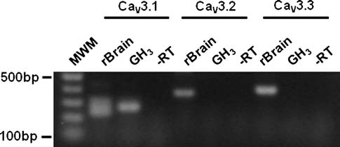
Standard RT-PCR was then performed on total RNA to
examine what sub-types of CaV3 channels were expressed
To validate GH3 cells as a model, we initially characterized
in GH3 cells, using sequence-specific primers. As shown in
the macroscopic currents using Ba2? as charge carrier and
Fig. , a PCR fragment of the predicted size was detected
then investigated whether CaV3 (LVA) channel expression
using primers for the CaV3.1 sequence. This PCR fragment
in this cell line was under the control of GCs. Whole cell
co-migrated with the product amplified from rat brain and
patch-clamp recordings confirmed the expression of two
HEK-293 cells stably transfected with the CaV3.1 cDNA
types of Ca2? currents in the plasma membrane of these
(not shown), which served as a positive control. The
cells as described previously (Meza et al. Whendepolarizations were carried out from a holding potential(Vh) of -80 mV to -20 mV, the current was small andtended to inactivate during a 200-ms pulse. This currentflows mostly through LVA channels. Larger depolariza-tions to ?20 mV induced the opening of additional LVAchannels and also recruited HVA channels, resulting in alarger inward current that inactivates rapidly at first andthen more slowly. The slowly inactivating phase of thecurrent is carried almost exclusively by HVA channels.
We next investigated the regulation of these currents
after acute (10–15 min) or chronic (5–6 days) treatmentwith HC or Dex. Macroscopic Ba2? current (IBa) throughCa2? channels was evoked by depolarizing voltage steps
Fig. 1 Chronic treatment of GH3 cells with GCs decreases whole-cell
from a Vh of -80 mV and the maximum amplitude
Ca2? currents through LVA channels. Comparison of the percentage
obtained at -20 mV and ?20 mV (normalized by C
of peak Ba2? current (through Ca2? channels) densities at -20 mV
were compared between GH
and Ba2? current densities measured at the end of activating pulses to
3 cells in the control condition
?20 mV in cells exposed to 5 lM dexamethasone (Dex) or
and in cells treated with GCs. IBa densities after acute
hydrocortisol (HC) for 5–6 consecutive days. Data are given as
treatment were similar in control and non-treated cells
mean ± S.E.M. (n = 7–24 cells in each experimental condition)
Cell Mol Neurobiol
identity of these amplicons was confirmed by sequencing.
In contrast, no PCR products could be amplified from theGH3 cells RNA when primers for the CaV3.2 sequencewere used, though these primers did amplify productscorresponding to the predicted size of cDNAs from ratbrain CaV3.2 (Fig. ) and from HEK-293 cells stablyexpressing this type of channel (not shown). Likewise, theuse of primers for the CaV3.3 sequence did not result in theproduction of a specific product from GH3 cells. Theseresults corroborate previous reports showing that one of thethree isoforms (CaV3.1) is expressed in the GH3 cells(Glassmeier et al. ; Mudado et al. In contrast toour findings, Mudado et al. ) reported the expressionof CaV3.3 channels in the GH3 cells, however their PCRexperiments revealed the presence of multiple hybridiza-tion bands with the CaV3.3 oligonucleotides, and no PCRproducts were apparently sequenced.
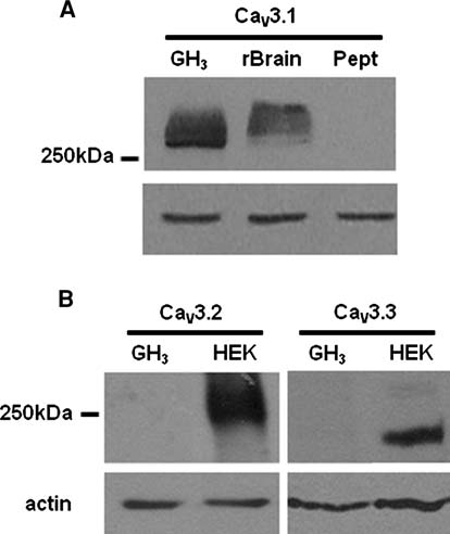
In order to investigate the expression of the LVA
channels at the level of protein, membranes derived fromGH3 cells were screened for the presence of CaV3 channelsusing specific antibodies. The results were concordant with
Fig. 3 CaV3 protein channel expression in GH3 cells. a CaV3.1
the RT-PCR analyses and showed that GH
protein expression was analyzed by western blot in GH3 or rat brain.
b Analysis of CaV3.2 and CaV3.3 proteins in GH3 cells or HEK-293
CaV3.1 but not CaV3.2 and CaV3.3 channels. As can be
cells stably expressing CaV3.2 and CaV3.3 channels. CaV proteins
seen in Fig. a, western blot analysis showed an immu-
were detected with specific polyclonal anti-CaV3 antibodies (Table )
noreactive protein with a molecular weight above 250 kDa,consistent with the predicted size of CaV3.1. Antibodyreactivity with the CaV3.1 protein from GH3 cells was
did recognize proteins corresponding to the predicted
compared with that of the protein expressed in the rat brain.
molecular weight in HEK-293 cells stably expressing the
Antibodies against CaV3.1 channels recognized the corre-
corresponding channels (Fig.
sponding expressed protein with similar molecular size.
Using a polyclonal antibody against the N-terminus of
The specificity of the CaV3.1 signal in GH3 cells was
CaV3.1 channel, we next studied whether chronic GC
evaluated using antibodies preadsorbed with its corre-
treatment (5–6 days) affected protein expression. Both
sponding antigenic synthetic peptide. There was no signal
plasma membrane and intracellular expression of the
for the CaV3.1 channels when the primary antibody was
CaV3.1 protein was analyzed. We first examined if the
preadsorbed (Fig. Likewise, no immunoreactivity
membrane localization of CaV3.1 channels was altered
could be found from GH3 cells when antibodies for CaV3.2
using a non-membrane permeant agent (sulfo-NHS-SS-
and CaV3.3 channels were used, although these antibodies
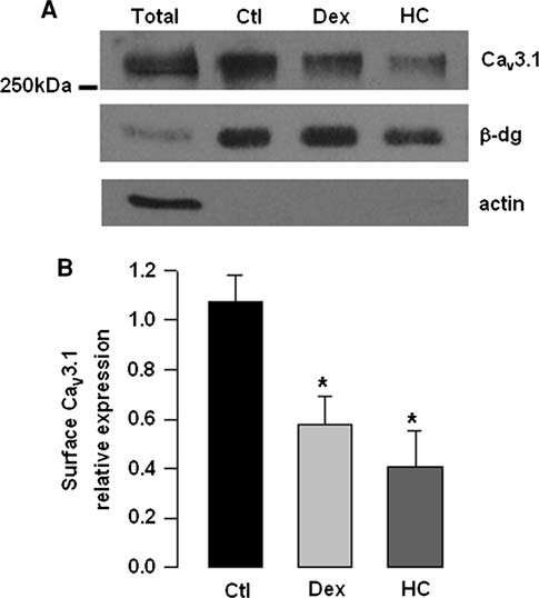
biotin) that biotinylates surface proteins and precipitatedthe proteins with avidin-linked streptavidin beads. Boundproteins were eluted and subjected to western blot analysis.
As shown in Fig. , the expression of biotinylated CaV3.1protein was significantly decreased in the GC treated cells.
The enrichment of cell membrane proteins in the biotin-ylated fractions and equal amounts of loaded proteins indifferent samples were confirmed by reprobing the blotswith an anti-b-dystroglycan antibody, a membrane marker.
Likewise, the biotinylated membrane fraction was notcontaminated by non-plasma membrane proteins becauseactin, an intracellular protein, was not detected in the
Fig. 2 CaV3 channel mRNA expression in GH3 cells. The presence
biotinylated fractions but was clearly detected in corre-
of CaV3 transcripts was probed by standard RT-PCR using cDNA
sponding cell lysates (total protein; Fig. a). These data
from rat brain as a positive control. MWM denotes molecular weights
suggested that chronic treatment with GCs reduces the
of the standards on the left. -RT indicates reactions run withoutreverse transcriptase
surface expression of CaV3.1 channels in GH3 cells.
Cell Mol Neurobiol
Fig. 4 Changes in CaV3.1 protein membrane expression in GH3 cellsinduced by chronic treatment with GCs. a Representative cell surfacebiotinylation assay using an anti-CaV3.1 specific antibody. b Densi-tometric quantification of three repetitions of the experiment shown in
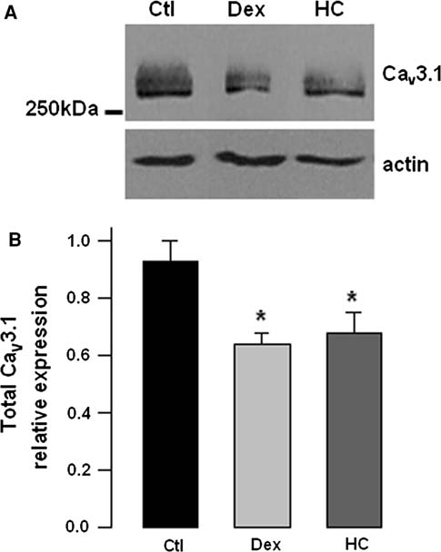
Fig. 5 Changes in CaV3.1 total protein expression by GCs in GH3
a. Asterisk denotes significant differences (P 0.05) between control
cells. a Total cell extracts from control and GC-treated cells were
and treated cells
analyzed by western-blot using an anti-CaV3.1 specific antibody.
Membranes were stripped and reprobed with an anti-actin antibody asa protein loading control. b Band signal intensities of CaV3.1 werenormalized to actin levels to obtain the relative expression of the
The preceding data suggested that the hormone treat-
CaV3.1 channels. Asterisk denotes significant differences (P 0.05)
ment may alter the trafficking of newly synthesized CaV3.1
between control and treated cells
proteins to the plasma membrane, resulting in a chronicdown-regulation of functional channel expression. There-
expression after GC treatment (Fig. ) and with the alter-
fore, we next quantified the relative levels of these proteins
ations in LVA current density (Fig. ).
in whole cell lysates by semi-quantitative western blot
The possibility exists, however, that GCs may be
experiments (Fig. ). The result of this analysis showed
affecting LVA currents by acting through a post-tran-
that the expression of the CaV3.1 protein was significantly
scriptional modification of the CaV3.1 channel protein. To
decreased to *60% of its control value in the cells treated
examine this, we compared the actions of Dex and HC on
with Dex and HC, which is consistent with the data
the whole-cell currents recorded in HEK-293 cells stably
obtained in the functional and biotinylation assays.
expressing CaV3.1 channels (Lee et al. Cribbs et al.
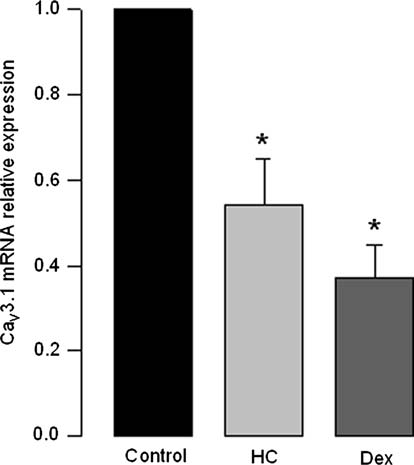
Taken as a whole, our observations suggested a tran-
). In this cell line, the expression of the channels is
scriptional mechanism by which GCs down-regulate
driven by the cytomegalovirus promoter (Cribbs et al.
CaV3.1 expression. To test this hypothesis, we next
), and therefore the transcriptional actions of the GCs
assessed the mRNA levels of this channel using quantita-
observed on native CaV3.1 channels may be bypassed.
tive PCR (qPCR). GH3 cells were treated 6 days with
Consequently, the post-transcriptional effects of the GCs
vehicle, Dex or HC (1 and 5 lM, respectively) before RNA
on the recombinant channels may be studied separately.
isolation. As expected from results obtained in RT-PCR
The results of this analysis show that the peak amplitude of
standard assays, using qPCR we detected transcripts of
the currents was very similar in both cell types (Supple-
CaV3.1 but not of CaV3.2 or CaV3.3 channels. More
mental Fig. 2). Likewise, the time course of the current
importantly, the result of these series of experiments
traces was almost identical in both experimental condi-
indicated that GC treatment caused a significant decrease
tions, suggesting that the activation and inactivation rates
(of *50%) in the mRNA levels for the CaV3.1 channels
of the CaV3.1 channels were not modified by the GC
(Fig. which is consistent with decreased channel protein
treatment. The steady-state inactivation properties of the
Cell Mol Neurobiol
is able to increase the rate of PRL gene transcription inGH3 cells (Day and Maurer Enyeart et al. ).
Likewise, it is well established that the expression levels
of LVA channels can be subjected to long-term regulationby different extracellular chemical messengers, including17b-oestradiol, insulin, neural growth factor, transforminggrowth factor-b1, and bone morphogenetic protein-2(Ritchie ; Meza et al. Avila et al. ; Lo´pez-Domı´nguez et al. Of particular interest is thatchronic treatment with GCs decreases LVA current densityin GH3 cells (Meza et al. ). Though the molecularbasis of this regulatory action is unknown, the slow onsetand reversion of this effect suggest a regulation at the levelof gene expression.
Indeed, decreased transcription of the cacna1g gene and
the consequent decrease in CaV3.1 channel protein mayaccount for the observed down-regulation of LVA channelsafter GCs treatment. Two sets of experimental evidencesupport this proposal. First, the quantitative PCR (qPCR)
Fig. 6 GCs treatment may affect CaV3.1 expression via transcrip-
analysis indicated that GCs treatment caused a significant
tional alterations. Quantitative PCR results of CaV3.1 mRNA levels
decrease (of *50%) in the mRNA levels for the CaV3.1
normalized to r18S RNA expression in control and treated GH3 cells.
Asterisk
channels, which is consistent with the reduction in channel
denotes significant differences (P 0.05) between control
and treated cells
protein expression after GCs treatment, and with thedecrease in LVA current density. Second, the gatingproperties of recombinant CaV3.1 channels heterologously
channels, characterized by applying a standard double-
expressed in HEK-293 were similar before and after GCs
pulse protocol, were also very similar between control and
treatment. In the HEK cells, the expression of the channels
treated cells (Supplemental Fig. 2).
is driven by the cytomegalovirus promoter and is thereforenot subject to the same transcriptional regulation thatcontrols native CaV3.1 channels in GH3 cells. Togetherthese data support the idea that the effects of GCs on
CaV3.1 channels are most likely mediated by decreasedtranscription.
Defining the function of voltage-gated Ca2? channels in
Based on our observations, we propose that this regu-
secretory and other cell types would be facilitated by the
lation is the result of alterations in gene expression that
identification of extracellular messengers, which preferen-
ultimately produce changes in the number of functional
tially regulate LVA channel functional expression. LVA
channels in the plasma membrane. Indeed, the most com-
channels are known to provide one of the major pathways
mon mechanism of action of GCs is the transcriptional
for Ca2? influx in GH3 clonal pituitary cells (Simasko et al.
regulation of specific target genes via GRs activation. After
; Herrington and Lingle Functional studies
translocation to the nucleus, GRs regulate transcription by
have shown that GH3 cells fire spontaneous Ca2?-depen-
binding to GREs of target genes. The rate of mRNA syn-
dent action potentials (SAPs; Scheru¨bl et al. and that,
thesis is either enhanced or repressed depending on the
whereas HVA channel activity is important for the
sequence of a given GRE and presumably on specific
upstroke of these SAPs, LVA channel activity is crucial in
interactions with other transcription factors (TFs).
regulating the discharge frequency (Llina´s and Yarom
For this reason, we searched for potential GREs in a
; Williams et al. ; LeBeau et al. Mansvel-
sequence that comprised *1.6 Kb of the genomic DNA
der and Kits ).
upstream of and including part of the first exon of the rat
Because the LVA channels regulate the firing of SAPs
cacna1g gene using the MatInspector software (Genomatix
and the electrical activity is coupled with hormone syn-
Software Inc; Ann Arbor, MI) and found numerous
thesis and secretion in GH3 cells (Schlegel et al. ), it is
TF-binding sites (for STAT1, 5 and 6; SMAD3 and 4;
reasonable to assume that these channels could be involved
NFjB and AP1) potentially relevant for GR signaling,
in GH and PRL production. Consistent with this, it has
upstream of the transcription initiation site (see Supple-
been shown that the influx of Ca2? through LVA channels
mental Fig. 3). It should be noted that this sequence has
Cell Mol Neurobiol
been cloned already and proved to be important during cell
differentiation (Bertolesi et al. ). Further studies areneeded to clarify the transcription factors that might be
Andrade A, Bermudez de Leon M, Hernandez-Hernandez O, Cisneros
B, Felix R (2007) Myotonic dystrophy CTG repeat expansion
mediating the GCs-induced regulation of the CaV3.1
alters Ca2? channel functional expression in PC12 cells. FEBS
Lett 581:4430–4438
What might be the biologic implications of such chronic
Avila T, Andrade A, Felix R (2006) Transforming growth factor-b1
GCs-mediated regulation of Ca
and bone morfogenetic protein-2 down-regulate Ca
v3.1 channels? Release of
expression in mouse C2C12 myoblasts. J Cell Physiol 209:448–
anterior pituitary hormones is regulated by diverse extra-
cellular messengers, including hypothalamic-hypophysio-
Bertolesi GE, Jollimore CA, Shi C, Elbaum L, Denovan-Wright EM,
tropic factors, hormones and growth factors. However, the
Barnes S, Kelly ME (2003) Regulation of a1G T-type calcium
mechanism by which these molecules act on target pitui-
channel gene (CACNA1G) expression during neuronal differ-entiation. Eur J NeuroSci 17:1802–1810
tary cells remains to be elucidated. Functional studies have
Catterall WA, Perez-Reyes E, Snutch TP, Striessnig J (2005)
shown that rat pituitary GH3 cells express both HVA and
International Union of Pharmacology. XLVIII. Nomenclature
LVA channels and that Ca2? current through LVA chan-
and structure-function relationships of voltage-gated calcium
nels is reduced by chronic treatment of the cells with GCs
channels. Pharmacol Rev 57:411–425
Chameau P, Qin Y, Spijker S, Smit G, Joels M (2007) Glucocorti-
(Meza et al. Hence, by diminishing the functional
coids specifically enhance L-type calcium current amplitude and
expression of LVA channels, GCs may induce a decrease
affect calcium channel subunit expression in the mouse hippo-
in the SAP firing frequency in GH3 cells. This action may
campus. J Neurophysiol 97:5–14
inhibit hormonal production at the level of transcription
Cribbs LL, Gomora JC, Daud AN, Lee JH, Perez-Reyes E (2000)
Molecular cloning and functional expression of Ca
and/or secretion.
T-type calcium channel from human brain. FEBS Lett 466:54–58
GCs have been implicated in attenuating plasma PRL
Day RN, Maurer RA (1990) Pituitary calcium channel modulation
in humans and abrogating PRL hypersecretion in rodents
and regulation of prolactin gene expression. Mol Endocrinol
(Piroli et al. ; Hubina et al. ). In GH
Enyeart JJ, Biagi B, Day RN (1990) Opposing actions of Bay K 8644
GCs also suppress PRL release elicited by various
enantiomers on calcium current, prolactin secretion, and synthe-
stimuli (Taylor et al. ). Likewise, elevated GCs
sis in pituitary cells. Mol Endocrinol 4:727–735
levels produce a marked impairment in somatic growth
Felix R (2005) Molecular regulation of voltage-gated Ca2? channels.
in both rodents and primates. It has been reported that
J Recept Signal Transduct Res 25:57–71
Fomina AF, Levitan ES, Takimoto K (1996) Dexamethasone rapidly
GCs play an important role in the regulation of GH
increases calcium channel subunit messenger RNA expression
synthesis and secretion, and it has been reported that
and high voltage-activated calcium current in clonal pituitary
cortisol inhibited growth hormone-releasing hormone
cells. Neuroscience 72:857–862
(GHRH)-stimulated GH release acting through a mech-
Garber SS, Hoshi T, Aldrich RW (1989) Regulation of ionic currents
in pheochromocytoma cells by nerve growth factor and dexa-
anism that involve Ca2? channels (Sartin et al. ;
methasone. J Neurosci 9:3976–3987
Watson et al. This is consistent with the fact that
Glassmeier G, Hauber M, Wulfsen I, Weinsberg F, Bauer CK,
in humans, GCs given chronically are associated with a
Schwarz JR (2001) Ca2? channels in clonal rat anterior pituitary
variety of negative side effects which limit the prolonged
cells (GH3/B6). Pflugers Arch 442:577–587
Gu Y, Preston MR, Magnay J, El Haj AJ, Publicover SJ (2001)
use of these hormones as therapeutic agents. Among
Hormonally-regulated expression of voltage-operated Ca2?
others, GCs treatment affects both the release and the
channels in osteocytic (MLO-Y4) cells. Biochem Biophys Res
effects of GH at the target sites, hence becoming func-
Commun 282:536–542
tional GH antagonists (Mauras
Hayashi T, Nakai T, Miyabo S (1991) Glucocorticoids increase Ca2?
uptake and [3H]dihydropyridine binding in A7r5 vascular
Although genomic mechanisms might be involved in
smooth muscle cells. Am J Physiol 261:C106–C114
mediating some of the responses detailed above, Ca2?
Herrington J, Lingle CJ (1992) Kinetic and pharmacological proper-
homeostasis may also play a significant role. Therefore,
ties of low voltage-activated Ca2? current in rat clonal (GH3)
more detailed studies on GCs chronic treatment linking
pituitary cells. J Neurophysiol 68:213–232
Hubina E, Nagy GM, Toth BE, Ivan G, Gorombey Z, Szabolcs I,
their regulatory actions on LVA channels to hormone
Kovacs L, Goth MI (2002) Dexamethasone and adrenocortico-
production and cell function are warranted.
tropin suppress prolactin secretion in humans. Endocrine18:215–219
This work was supported by funds from Cona-
Joels M, Velzing E, Nair S, Verkuyl JM, Karst H (2003) Acute stress
cyt to RF. We thank Drs. M. E. Mendoza (Cinvestav-IPN, Mexico)
increases calcium current amplitude in rat hippocampus:
and J. C. Gomora (IFC-UNAM, Mexico) for the generous gift of the
temporal changes in physiology and gene expression. Eur J
cell lines and Dr. M. Hernandez (Cinvestav-IPN, Mexico) and
NeuroSci 18:1315–1324
D. Mornet (INSERM ERI 25 Muscle et Pathologies, France) for the
Karst H, Wadman WJ, Joels M (1994) Corticosteroid receptor-
anti-actin and anti-b-dystroglycan antibodies, respectively. We are
dependent modulation of calcium currents in rat hippocampal
also indebted to J. Arikkath for critically reviewing the manuscript.
CA1 neurons. Brain Res 649:234–242
Cell Mol Neurobiol
Kassel O, Herrlich P (2007) Crosstalk between the glucocorticoid
glucocorticoid receptors and glucocorticoid negative feedback
receptor and other transcription factors: molecular aspects. Mol
on prolactin secretion in estrogen-induced pituitary tumors.
Cell Endocrinol 275:13–29
Kato H, Hayashi T, Koshino Y, Kutsumi Y, Nakai T, Miyabo S
Publicover SJ, Thomas GP, el Haj AJ (1994) Induction of a low
(1992) Glucocorticoids increase Ca2? influx through dihydro-
voltage-activated, fast-inactivating Ca2? channel in cultured
pyridine-sensitive channels linked to activation of protein kinase
bone marrow stromal cells by dexamethasone. Calcif Tissue Int
C in vascular smooth muscle cells. Biochem Biophys Res
Commun 188:934–941
Ritchie AK (1993) Estrogen increases low voltage-activated calcium
LeBeau AP, Robson AB, McKinnon AE, Donald RA, Sneyd J (1997)
current density in GH3 anterior pituitary cells. Endocrinology
Generation of action potentials in a mathematical model of
corticotrophs. Biophys J 73:1263–1275
Sartin JL, Kemppainen RJ, Coleman ES, Steele B, Williams JC
Lee JH, Daud AN, Cribbs LL, Lacerda AE, Pereverzev A, Klockner
(1994) Cortisol inhibition of growth hormone-releasing hor-
U, Schneider T, Perez-Reyes E (1999) Cloning and expression of
mone-stimulated growth hormone release from cultured sheep
a novel member of the low voltage-activated T-type calcium
pituitary cells. J Endocrinol 141:517–525
channel family. J Neurosci 19:1912–1921
Scheru¨bl H, Hescheler J, Bychkov R, Cuber JC, John M, Riecken EO,
Llina´s R, Yarom Y (1981) Properties and distribution of ionic
Wiedenmann B (1994) Electrical activity and calcium channels
conductances generating electroresponsiveness of mammalian
in neuroendocrine cells. Ann NY Acad Sci 733:335–339
inferior olivary neurons in vitro. J Physiol 315:569–584
Schlegel W, Winiger BP, Mollard P, Vacher P, Wuarin F, Zahnd GR,
Lo´pez-Domı´nguez AM, Espinosa JL, Navarrete A, Avila G, Cota G
Wollheim CB, Dufy B (1987) Oscillations of cytosolic Ca2? in
(2006) Nerve growth factor affects Ca2? currents via the p75
pituitary cells due to action potentials. Nature 329:719–721
receptor to enhance prolactin mRNA levels in GH3 rat pituitary
Simasko SM, Weiland GA, Oswald RE (1988) Pharmacological
cells. J Physiol 574(Pt 2):349–365
characterization of two calcium currents in GH3 cells. Am J
Mansvelder HD, Kits KS (2000) All classes of calcium channel
Physiol 254(3 Pt 1):E328–E336
couple with equal efficiency to exocytosis in rat melanotropes,
Takimoto K, Li D, Nerbonne JM, Levitan ES (1997) Distribution,
inducing linear stimulus-secretion coupling. J Physiol 526(Pt
splicing and glucocorticoid-induced expression of cardiac a1C
and a1D voltage-gated Ca2? channel mRNAs. J Mol Cell Cardiol
Mauras N (2001) Growth hormone therapy in the glucocorticosteroid-
dependent child: metabolic and linear growth effects. Horm Res
Taylor AD, Cowell AM, Flower RJ, Buckingham JC (1995)
56(Suppl 1):13–18
Dexamethasone suppresses the release of prolactin from the rat
Meza U, Avila G, Felix R, Gomora JC, Cota G (1994) Long-term
anterior pituitary gland by lipocortin-1 dependent and indepen-
regulation of calcium channels in clonal pituitary cells by
dent mechanisms. Neuroendocrinology 62:530–542
epidermal growth factor, insulin, and glucocorticoids. J Gen
Wang L, Feng ZP, Duff HJ (1999) Glucocorticoid regulation of
Physiol 104:1019–1038
cardiac K? currents and L-type Ca2? current in neonatal mice.
Mudado MA, Rodrigues AL, Prado VF, Beirao PS, Cruz JS (2004)
Circ Res 85:168–173
CaV3.1 and CaV3.3 account for T-type Ca2? current in GH3
Watson S, Porter RJ, Young AH (2000) Effect of hydrocortisone on
cells. Braz J Med Biol Res 37:929–935
the pituitary response to growth hormone releasing hormone.
Naess O, Haug E, Gautvik K (1980) Effects of glucocorticosteroids
Psychopharmacology (Berl) 152:40–46
on prolactin and growth hormone production and characteriza-
Williams PJ, MacVicar BA, Pittman QJ (1990) Electrophysiological
tion of the intracellular hormone receptors in rat pituitary tumour
properties of neuroendocrine cells of the intact rat pars
cells. Acta Endocrinol (Copenh) 95:319–327
intermedia: multiple calcium currents. J Neurosci 10:748–756
Obejero-Paz CA, Lakshmanan M, Jones SW, Scarpa A (1993) Effects
Zhou J, Cidlowski JA (2005) The human glucocorticoid receptor: one
of dexamethasone on L-type calcium currents in the A7r5
gene, multiple proteins and diverse responses. Steroids 70:407–
smooth muscle-derived cell line. FEBS Lett 333:73–77
Piroli G, Grillo C, Ferrini M, Diaztorga G, Rey E, Libertun C,
Source: http://www.fisiologia.buap.mx/laboratorios/sensorial/pdf/2009%20Traudy%20et%20al.,%20Cell%20Mol%20Neurobiol_2009.pdf
Pharmaceutical Applications Compendium PURITY AND IMPURITY ANALYSIS APPLICATION OF CHROMATOGRAPHY AND MASS SPECTROMETRY IN PHARMACEUTICAL PURITY AND IMPURITY ANALYSIS Accurate assessment of product quality requires robust analytical methods. To remain competitive, drug development and quality control programs must find efficient and effective ways to develop new analytical methods and to transfer and optimize existing procedures. Increased regulatory attention on the control of impurities (e.g., much lower thresholds for genotoxic impurities) has fueled the need for analytical procedures that al ow significantly lower detection limits. This in turn requires more sensitive instruments and places higher demands on selectivity, since many additional impurities may be present at lower concentration ranges. UHPLC offers significant advantages in analytical performance, speed, sensitivity, and resolution. Today's labs must also col aborate with global partners using a variety of instruments. As a result, flexibility in method development and transfer has become a key component of success in the pharmaceutical industry. The application of various mass spectroscopy (MS) detectors to genotoxic impurity analysis has enabled breakthrough detection limits of as little as a few hundred ppm. MS-based methods general y provide additional robustness compared to techniques such as UV alone, due to their high specificity and sensitivity.
You will find on the followingpages all the necessary informa-tion for a correct use of theworking unit. Therefore we sug-gest that the user carefully readsand follows the technical instruc-tions. Moreover we advise tokeep the user guide in a safe andhandy place. The above mentio-ned guide can be subjected tomodifications and further impro-vements. GENERAL DESCRIPTION