Nara-wu.ac.jp

Brain 2011: 134; 892–902 A JOURNAL OF NEUROLOGY The clinical and molecular genetic featuresof idiopathic infantile periodic alternatingnystagmus Mervyn G. Thomas,1 Moira Crosier,2 Susan Lindsay,2 Anil Kumar,1 Shery Thomas,1Masasuke Araki,3 Chris J. Talbot,4 Rebecca J. McLean,1 Mylvaganam Surendran,1 Katie Taylor,5Bart P. Leroy,6 Anthony T. Moore,7,8 David G. Hunter,9 Richard W. Hertle,10,11 Patrick Tarpey,12Andrea Langmann,13 Susanne Lindner,13 Martina Brandner13 and Irene Gottlob1 1 Ophthalmology Group, School of Medicine, University of Leicester, RKCSB, PO Box 65, Leicester LE2 7LX, UK2 MRC-Wellcome Trust Human Developmental Biology Resource (Newcastle), Institute of Human Genetics, Newcastle University, International Centre for Life, Newcastle upon Tyne NE1 3BZ, UK 3 Department of Biological Sciences, Developmental Neurobiology Laboratory, Nara Women's University, Nara 630-8506, Japan4 Department of Genetics, University of Leicester, University Road, Leicester LE1 7RH, UK5 Department of Cancer Studies and Molecular Medicine, University of Leicester, RKCSB, Leicester LE2 7LX, UK6 Department of Ophthalmology and Centre for Medical Genetics, Ghent University and Ghent University Hospital, 9000 Ghent, Belgium 7 Moorfields Eye Hospital, City Road, London EC1V 2PD, UK8 UCL Institute of Ophthalmology, London EC1V 9EL, UK9 Department of Ophthalmology, Children's Hospital Boston, Harvard Medical School, Boston, MA 02115, USA 10 Department of Ophthalmology, Children's Hospital Medical Centre of Akron, Akron, OH 44308, USA11 Northeast Ohio College of Medicine, Rootstown, OH 44272, USA12 Wellcome Trust Sanger Institute, Hinxton Cambridge CB10 1SA, UK13 Department of Ophthalmology, Medical University Graz, Auenbruggerplatz 4, 8036, Graz, Austria Correspondence to: Prof. Irene Gottlob,Ophthalmology Group,School of Medicine,University of Leicester,RKCSB, PO Box 65,Leicester LE2 7LX,UKE-mail: [email protected] Periodic alternating nystagmus consists of involuntary oscillations of the eyes with cyclical changes of nystagmus direction.
It can occur during infancy (e.g. idiopathic infantile periodic alternating nystagmus) or later in life. Acquired forms are oftenassociated with cerebellar dysfunction arising due to instability of the optokinetic-vestibular systems. Idiopathic infantile peri-odic alternating nystagmus can be familial or occur in isolation; however, very little is known about the clinical characteristics,genetic aetiology and neural substrates involved. Five loci (NYS1-5) have been identified for idiopathic infantile nystagmus;three are autosomal (NYS2, NYS3 and NYS4) and two are X-chromosomal (NYS1 and NYS5). We previously identified theFRMD7 gene on chromosome Xq26 (NYS1 locus); mutations of FRMD7 are causative of idiopathic infantile nystagmus influen-cing neuronal outgrowth and development. It is unclear whether the periodic alternating nystagmus phenotype is linked toNYS1, NYS5 (Xp11.4-p11.3) or a separate locus. From a cohort of 31 X-linked families and 14 singletons (70 patients) withidiopathic infantile nystagmus we identified 10 families and one singleton (21 patients) with periodic alternating nystagmus ofwhich we describe clinical phenotype, genetic aetiology and neural substrates involved. Periodic alternating nystagmus was not Received September 3, 2010. Revised November 8, 2010. Accepted November 9, 2010. Advance Access publication February 8, 2011ß The Author (2011). Published by Oxford University Press on behalf of the Guarantors of Brain. All rights reserved.
For Permissions, please email: [email protected] Idiopathic infantile PAN Brain 2011: 134; 892–902 detected clinically but only on eye movement recordings. The cycle duration varied from 90 to 280 s. Optokinetic reflex was notdetectable horizontally. Mutations of the FRMD7 gene were found in all 10 families and the singleton (including three novelmutations). Periodic alternating nystagmus was predominantly associated with missense mutations within the FERM domain.
There was significant sibship clustering of the phenotype although in some families not all affected members had periodicalternating nystagmus. In situ hybridization studies during mid-late human embryonic stages in normal tissue showed restrictedFRMD7 expression in neuronal tissue with strong hybridization signals within the afferent arms of the vestibulo-ocular reflexconsisting of the otic vesicle, cranial nerve VIII and vestibular ganglia. Similarly within the afferent arm of the optokinetic reflexwe showed expression in the developing neural retina and ventricular zone of the optic stalk. Strong FRMD7 expression wasseen in rhombomeres 1 to 4, which give rise to the cerebellum and the common integrator site for both these reflexes (vestibularnuclei). Based on the expression and phenotypic data, we hypothesize that periodic alternating nystagmus arises from instabilityof the optokinetic-vestibular systems. This study shows for the first time that mutations in FRMD7 can cause idiopathic infantileperiodic alternating nystagmus and may affect neuronal circuits that have been implicated in acquired forms.
Keywords: periodic alternating nystagmus; FRMD7; optokinetic reflex; vestibulo-ocular reflex; in situ hybridizationAbbreviation: PAN = periodic alternating nystagmus form of PAN often responds well to specific drugs such as baclo- fen (Halmagyi et al., 1980).
Nystagmus is defined as the involuntary rhythmic oscillations of Idiopathic infantile nystagmus is a genetically heterogeneous the eyes, with a reported prevalence of approximately 2.4 in 1000 condition with X-linked, autosomal dominant and autosomal re- (Sarvananthan et al., 2009). There is significant negative social cessive modes of inheritance reported. To date, five chromosomal stigma and relatively poor visual function scores reported with loci (NYS 1–5) have been described in the literature associated this condition (Pilling et al., 2005). In infantile nystagmus, these with idiopathic infantile nystagmus (Table 1). Within the NYS1 oscillations are horizontal and conjugate and are characterized by locus (Xq26.2), the FRMD7 gene was identified, mutations of two components: (i) a slow drift of the eyes (called the slow which cause X-linked idiopathic infantile nystagmus (Tarpey phase); followed by (ii) a corrective fast eye movement (called et al., 2006; Schorderet et al., 2007; Zhang et al., 2007).
the quick phase) that is responsible for realigning the fovea to Previous studies have shown that the penetrance among female the object of interest. In idiopathic infantile periodic alternating carriers is 50% (Tarpey et al., 2006; Thomas et al., 2008).
nystagmus (PAN), the direction of the quick phase and slow Expression of FRMD7 has been shown in neuronal tissue in the phase alternates periodically with time. This phenotype of nystag- developing retina, mid and hind brain (Tarpey et al., 2006; mus is distinct from other nystagmus forms due to the periodic Betts-Henderson et al., 2009), although it is not clear which spe- time component. Acquired forms of PAN are also reported and cific gaze control systems are affected by mutations in the gene.
arise due to instability of the vestibulo-optokinetic systems (Leigh Unaffected female carriers can have a subnormal optokinetic nys- et al., 1981). Animal and mathematical models for acquired PAN tagmus gain (Thomas et al., 2008). Recent studies in neuro-2A have demonstrated how instability of the velocity storage mech- cells have demonstrated a role for FRMD7 in neuronal outgrowth anism for vestibular eye movements and an adaptive mechanism and development (Betts-Henderson et al., 2009).
for this instability can result in a periodicity of oscillations of 4 min Idiopathic infantile PAN is considered to be a subtype of idio- (Waespe et al., 1985; Leigh and Khanna, 2006). It has also been pathic infantile nystagmus; however, its diagnosis has different shown that patients with acquired PAN have abnormalities of optokinetic nystagmus, with some patients having no optokinetic (Reinecke, 1997). The first report of familial idiopathic infantile nystagmus response and the PAN cycle continues through the PAN was by Huygen and colleagues (1995), where both mother optokinetic nystagmus stimuli (Baloh et al., 1976). The acquired Table 1 The nystagmus loci (Tarpey et al., 2006) Autosomal dominant (Kerrison et al., 1996) Autosomal dominant (Klein et al., 1998) Autosomal dominant (Ragge et al., 2003) NYS 5 (Xp11.4-p11.3) (Cabot et al., 1999) = Online Mendelian Inheritance in Man.
Brain 2011: 134; 892–902 M. G. Thomas et al.
Shallo-Hoffmann et al. (1999, 2004) reported a case of idiopathic consisted of square-wave contrast gratings of 2.2 cycle size and infantile PAN with an X-linked history of nystagmus. Hertle et al.
Michelson contrast 0.88 cd/m2. Optokinetic nystagmus was tested at (2005) described another family spanning three generations, with 20/s velocity in both horizontal (stimulus direction: rightwards and a phenotype of PAN in all four patients examined with eye move- leftwards) and vertical directions (upwards and downwards). For fur-ther details of the experimental setup see Thomas et al. (2008).
ment recordings. Based on an X-linked mode of inheritance and Informed consent was obtained from all subjects participating in this the distinct phenotype consistently seen in all generations it was study. The study adhered to the tenets of the Declaration of Helsinki assumed that a unique locus for idiopathic infantile PAN was and was approved by the local Ethics Committee.
present on the X-chromosome. However, no molecular geneticstudies were performed in these families to substantiate that aseparate locus is present for idiopathic infantile PAN. There is Sequencing and mutation analysis some evidence for an additional locus for idiopathic infantile nys- Bidirectional sequence analysis of the exons and intron/exon junctions tagmus on the X-chromosome. Cabot et al. (1999) reported a four of the FRMD7 gene were performed using DNA from the proband generation French family with X-linked idiopathic infantile nystag- within each of the new families and singletons with idiopathic infantile mus. Linkage analysis showed mapping to Xp11.4-11.3 between nystagmus where no genetic test for FRMD7 had been performed the polymorphic markers DXS8015 and DXS1003, however, these previously. Primer details, expected product sizes and annealing tem- investigators did not describe the type of nystagmus in this family.
perature are shown in Table 2. Mutation analysis software Seqscape Due to the limited phenotypic data in the literature it is currently version 2.1.1 (Applied Biosystems) was used for base calling and align- unknown whether idiopathic infantile PAN occurs in nystagmus ment of the contigs. The Genbank file (NM_194277.2) was imported with other inheritance patterns. In the family described by into Seqscape and used as the reference complementary DNA se- Huygen and colleagues (1995), the inheritance pattern is not quence for contig alignment. Base position + 1 corresponded to A ofthe translation initiation codon ATG. Intronic sequence changes were clear. In this study we present evidence showing that mutations identified based on the FRMD7 genomic sequence (NC_000023.10) of the FRMD7 gene can be associated with PAN. We report three and amino acid changes were identified based on the reference protein novel mutations and eight previously described mutations of the sequence (NP_919253.1). Allelic variations were assessed against the FRMD7 gene associated with PAN. Furthermore, we highlight the sequence data from 300 male controls (without nystagmus).
spectrum of variable phenotypes associated with FRMD7 muta-tions and present evidence that expression of FRMD7 correlates to neuronal circuits that have also been associated with acquired In situ hybridization experiments Human embryonic and foetal tissues were obtained from theMRC-Wellcome Trust Human Developmental (www.hdbr.org; Lindsay and Copp, 2005), Institute of Human Materials and methods Genetics, Newcastle University. The samples were collected with ap-propriate maternal consents and ethical approval by the Newcastle and Patients and clinical examinations North Tyneside Research Ethics Committee. Tissue sections used em-bryos with a normal karyotype and morphology. Tissue sections from Using eye movement recordings we examined a cohort of 31 families eight samples were analysed from Carnegie Stage 16 (37 days post- with X-linked idiopathic infantile nystagmus and 14 singletons (total of conception; n = 1); Carnegie Stage 19 (47 days post-conception; 70 patients). Fifteen of the families were previously described to have n = 3); Carnegie Stage 22 (54 days post-conception; n = 2) and FRMD7 mutations (Tarpey et al., 2006) and four families did not have Carnegie Stage 23 (56 days post-conception; n = 2). The stage of FRMD7 mutations. For the remaining 12 families with idiopathic embryonic development was determined by assessment of external infantile nystagmus, the genotype was not yet determined. Among morphology as described in Bullen and Wilson (1997) and O'Rahilly the singletons, we included the previously identified three singletons and Muller (1987).
with FRMD7 mutations (Tarpey et al., 2006) and 11 singletons with In situ hybridizations were preformed as previously described idiopathic infantile nystagmus that were not yet sequenced. Within (Moorman et al., 2001) with some modifications. Briefly, sections this cohort we identified 10 families (Fig. 1) and one singleton with were dewaxed in xylene, gradually hydrated in decreasing ethanol idiopathic infantile PAN (total of 21 patients). In our study population, concentrations before incubation in proteinase K (20 mg/ml) at room idiopathic infantile PAN occurred in 18 males and in three females.
temperature, followed by fixation using 4% paraformaldehyde in Detailed ophthalmic examination was performed in all patients. At phosphate buffered saline. Background was reduced by treating with least one affected patient within each family underwent electrodiag- 0.1 M triethanolamine pH 8. Sections were air dried and the sense or nostic examinations according to the International Society for Clinical antisense probes [300 ng labelled probe per 100 ml of DIG Easy Hyb Electrophysiology of Vision standards (visually evoked potentials and mix (Roche)] were added for hybridization at 68C overnight. Next electroretinography) to exclude other infantile forms of nystagmus day sections were washed in 5 followed by 2 saline sodium citrate such as those associated with albinism or retinal diseases. Eye move- buffer at 60C then incubated with anti-digoxigenin alkaline phosphat- ment recordings were performed (EyeLink II, 500 Hz, SR Research, ase Fab fragments (Roche) diluted 1:1000 at 4C overnight. Sections Toronto, Canada) using a central fixation task over a prolonged were then washed and expression detected using NBT/BCIP (20 ml/ml; period of 5 min to detect idiopathic infantile PAN. Several members Roche) in 0.1 M Tris (pH 9.5)/0.1 M NaCl (Buffer 2) in the dark at of most families and all the singletons underwent the prolonged fix- room temperature. Developing was stopped by rinsing slides in Buffer ation task (Fig. 1).
2 then distilled water. Sections were mounted using Aquamount and In addition, optokinetic nystagmus was tested in all affected patients analysed using a Zeiss Axioplan 2 microscope. Images were captured with idiopathic infantile PAN. The optokinetic nystagmus stimulus with Zeiss Axiovision 4 imaging system.
Idiopathic infantile PAN Brain 2011: 134; 892–902 Figure 1 Families with idiopathic infantile PAN. In Families 1–4 eye movement recordings were performed in three or more affectedindividuals, whereas in Families 4–7 eye movement recordings were obtained in two or more affected individuals. In Families 8–10 eyemovement recordings were only performed in one affected individual.
0.2 LogMAR. Three patients with idiopathic infantile PAN had aslight anomalous head posture between 5–10. However, in onlyone patient was the head posture noticed to alternate to the right Clinical characteristics and eye or left on two different examination days. None of the patients movement abnormalities with idiopathic infantile PAN had strabismus. All had some degreeof stereopsis; the range of stereoacuity was 85–55000 with a The pedigrees of patients diagnosed with idiopathic infantile PAN median stereoacuity of 15000.
are shown in Fig. 1. Among the 10 families, we were able to Idiopathic infantile PAN was not diagnosed clinically in any of perform eye movement recordings in 26 affected patients, of the families or the singletons. However, the use of eye movement whom 20 had idiopathic infantile PAN (Fig. 1). The phenotypes recordings during a prolonged duration of fixation (5 min) aided in affected members of Family 2 have previously been described the diagnosis of idiopathic infantile PAN. An example of original by Hertle et al. (2005).
eye movement recordings from Family F3 is shown in Fig. 2. In The best-corrected visual acuity in our cohort of patients with Families F1, F3, F4 and F7 we observed phenotypic heterogeneity PAN ranged from 0.0 LogMAR to 0.54 LogMAR with a median of as not all the examined patients had idiopathic infantile Brain 2011: 134; 892–902 M. G. Thomas et al.
Table 2 Forward and reverse primer sequences, product families; similarly within the 11 singletons we identified mutations sizes and melting temperature (Tm) used to amplify in two out of 11 singletons. Thus, the total cohort consisted of a total of 24 families and five singletons with an FRMD7 mutation;the remaining seven families and nine singletons had no such mu- tations. In all 10 of the idiopathic infantile PAN families and the singletons with idiopathic infantile PAN, there were mutations ofthe FRMD7 gene, three of which were novel. A summary of the mutations and domains affected are shown in Fig. 4.
The three novel mutations were in Family F8, F9 and singleton S1. Family F8 had a missense mutation (c.47T 4 C) in exon 1, resulting in the substitution of phenylalanine by serine at position 16 (p.F16S). Sequence analysis of the proband in family F9 re- vealed a splice-site mutation (c.58-1G 4 A) at the 30-end of intron 1. This results in the loss of the conserved splice acceptor residue. The effects of the mutation were predicted using the al- ternative splice-site predictor (Wang and Marı´n, 2006), and con- sidered pathological due to exon skipping resulting in a messenger RNA transcript with exon 2 missing. The singleton S1, had a mis- sense mutation (c.811T 4 A) in exon 9 resulting in a substitution of cysteine to serine at position 271 (p.C271S). Both missense mutations at amino acid positions 16 and 271 were considered pathological as they involved residues that were identical within invariant blocks in the species Mus, Gallus, Xenopus and Tetraodon. Furthermore, the structural effects of these mutations were elucidated based on the known crystal structure of the clo- sest orthologue (PDB Accession ID: 1GG3) and secondary struc- ture prediction of the FRMD7 protein sequence using Emboss Garnier (Garnier et al., 1978). Finally the stability of the mutant protein was assessed using I-Mutant (v2.0) (Capriotti et al., 2005) and Coot (v0.6) (Emsley and Cowtan, 2004). The amino acid change C271S would disrupt a large alpha-helical domain in the wild-type structure. Similarly, the amino acid change F16S is likely to disrupt adjacent secondary structure. Consequently, both mis-sense mutations, F16S and C271S, decrease the stability of themutant protein (due to a decrease in the free energy value).
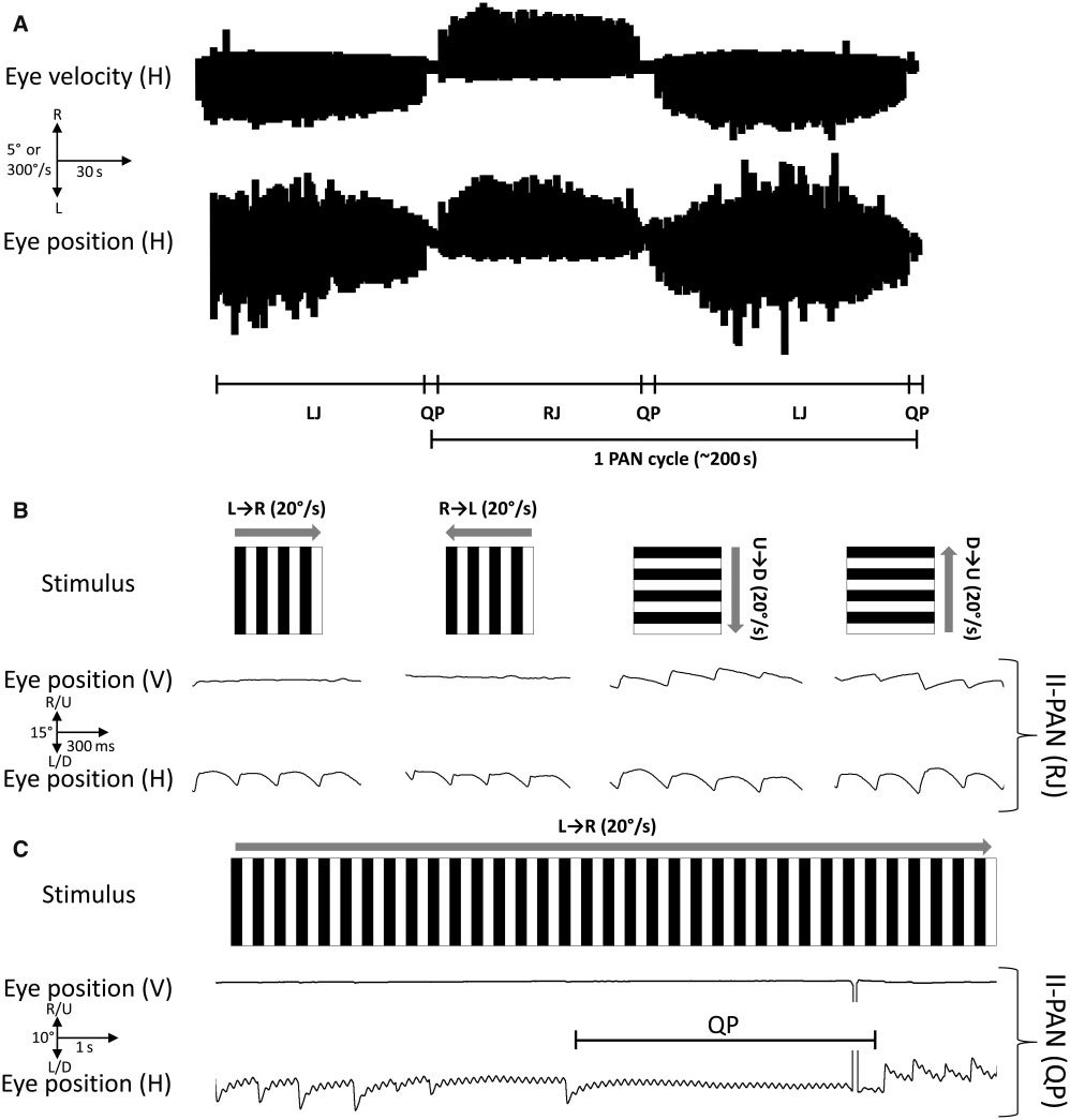
Six of the eight remaining families (F1, 2, 3, 4, 7 and 10) all PAN (Fig. 2). Among the families with idiopathic infantile PAN, six revealed missense mutations of the FRMD7 gene. These mutations of 26 subjects in which eye movements were performed did not have been reported previously and their translational effects have have PAN. Overall, the time period for the idiopathic infantile PAN been described (Tarpey et al., 2006). Family F5 and F6 were the cycle varied between 90 and 260 s and the singleton had a peri- only families that had a nonsense mutation (c. 1003C 4 T) result- odicity of 280 s. All family (F1–F10) members with idiopathic in- ing in a premature stop codon at amino acid position 335 (p.
fantile PAN had a jerk-related or dual jerk nystagmus. In family R335X). Family F2 was previously reported with an idiopathic in- members without idiopathic infantile PAN (F1, III:3; F3, II:2 and 4; fantile PAN phenotype and an X-linked inheritance (Hertle et al., F4, III:1 and 2; F7, II:1;) the predominant nystagmus waveform 2005). Sequence analysis of this family revealed a missense mu- was pendular.
tation (c.70G 4 A) resulting in hemizygous replacement of glycine None of the patients with idiopathic infantile PAN showed an with arginine at position 24 (p. G24R). The amino acid changes as optokinetic response for either horizontal stimulus directions a result of the missense mutations in families F1 (L231V), F2 (rightwards and leftwards). The PAN cycle continued through (G24R), F3 (C271Y), F4 (A266P), F7 (A226T) and F10 (S340L) the optokinetic nystagmus testing and was not changed by the are associated with a decrease in the free energy value that is optokinetic nystagmus stimuli (Fig. 3). Vertical optokinetic nystag- likely to destabilize the mutant protein.
mus was seen in all patients in both directions (upwards anddownwards).
Expression of FRMD7 In situ hybridization experiments showed strong hybridization sig-nals from the structures involved in setting up the vestibulo-ocular Among the 12 families with idiopathic infantile nystagmus we reflex and optokinetic reflex arc, this included the developing identified mutations in nine out of 12 previously uncharacterized Idiopathic infantile PAN Brain 2011: 134; 892–902 Figure 2 Original eye movement recordings from Family F1. Overviews of the three phases of the PAN cycle and excerpts from withineach phase of the cycle are shown in this figure. A typical PAN cycle consists of three phases: (i) left jerk (LJ), where the quick phase isdirected to the left; (ii) a quiet phase (QP), where the intensity of the nystagmus is minimal; and (iii) a right jerk (RJ), where the quick phaseis directed to the right. One of the examined family members (III:3) did not have PAN, but a pendular (P) nystagmus. Scale for the excerptsare shown in the bottom right with waveform deflection upwards and downwards representing horizontal eye movements to the right andleft, respectively.
retina (Fig. 5A–E). Expression is detected in the ventricular zone of recordings and most patients had relatively good visual acuity the neural retina and optic stalk (Fig. 5B). In structures of the (median: 0.2 LogMAR) and stereoacuity (median: 15000). Fewer vestibulo-ocular reflex, FRMD7 is expressed in the otic vesicle and females were affected since FRMD7-related infantile nystagmus vestibulocochlear ganglion (Fig. 5C). The vestibular nuclei, which represents a disorder associated with variable penetrance in fe- arise partly from the ventricular zone of rhombomeres 2, 3 and 4, males (Tarpey et al., 2006; Thomas et al., 2008). The PAN cycle form the horizontal neural integrator, which is an important structure length varied from 90–280 s and none of the patients with idio- in the vestibulo-ocular reflex and optokinetic reflex arcs. FRMD7 pathic infantile PAN had a horizontal optokinetic reflex. We show expression is seen in the ventricular zone of rhombomeres 2, 3 and that FRMD7 is expressed within developing vestibulo-ocular reflex 4 (Fig. 5D) as well as in the developing cerebellum (Fig. 5E). The and optokinetic reflex arcs, which identifies the likely neural sub- cerebellum arises entirely from rhombomere 1 and its ventricular strates involved in idiopathic infantile PAN and in FRMD7-related zone gives rise to neuroblasts that migrate on radial glia to develop infantile nystagmus. We identified 11 mutations in 10 families and into the cerebellar nuclei and Purkinje cells in the cerebellar cortex.
one singleton and describe both the phenotypic and translational FRMD7 is expressed in differentiating and migrating neurons as well effects of these mutations. In our cohort of families, the predom- as in the ventricular zone, for example in the cerebellum and rhom- inant class of mutation associated with this phenotype were mis- bomeres 3 and 4 in the hindbrain (Fig. 5D and E) but also in the sense mutations (8/11) though both truncating (2/11) and subpallium in the forebrain (data not shown).
splice-site (1/11) mutations are also seen. Ten of the 11 mutationsresulted in amino acid changes within functionally significant do- mains (FERM-N, FERM-C and FA).
From a clinical point of view PAN is typically under-diagnosed In this study we show for the first time that idiopathic infantile (Abadi and Pascal, 1994; Gradstein et al., 1997; Abadi and Bjerre, PAN can be associated with mutations of the FRMD7 gene.
2002) as it can often only be identified on eye movement record- Idiopathic infantile PAN was only detected using eye movement ings during an extended fixation task to demonstrate the


Brain 2011: 134; 892–902 M. G. Thomas et al.
Figure 3 Compressed eye movement recordings showing an overview of the various phases of the PAN cycle (A). In the above exampleone cycle consists of right jerk (RJ) followed by a quiet phase (QP), left jerk phase (LJ) and another quiet phase (QP). Upward deflection ofthe horizontal (H) position and velocity trace represents right-beating nystagmus and downward deflection represents left-beating nys-tagmus. The optokinetic response was measured for optokinetic nystagmus stimuli (B) moving in the horizontal [rightwards (L!R) andleftwards (R!L)] and in the vertical direction [downwards (U!D) and upwards (D!U)]. The patient (II-PAN) shows no horizontaloptokinetic response to the stimulus; the nystagmus is unchanged in the right jerk phase during optokinetic nystagmus testing. Howeverfor the vertical optokinetic nystagmus stimuli, a vertical optokinetic response is seen in the vertical trace (V) for the patient. The idiopathicinfantile PAN cycle was not changed by the horizontal optokinetic nystagmus stimuli as shown in (C). The transition (QP) between left jerkand right jerk is seen during an extended horizontal optokinetic nystagmus task.
periodicity of the nystagmus and its three phases. A recent study nystagmus patients have PAN. Interestingly, in the idiopathic in- estimated that 15% of all infantile nystagmus syndrome patients fantile PAN incidence study by Shallo-Hoffmann and colleagues have PAN (Hertle et al., 2009). In contrast, Shallo-Hoffmann et al.
(1999, 2004), a family with X-linked PAN was described though (1999) showed that a higher proportion of 39% of congenital no genetic diagnosis was provided. We noticed the occurrence of Idiopathic infantile PAN Brain 2011: 134; 892–902 Figure 4 Country of origin, mutations of the FRMD7 gene in the families (F1–10) and singleton (S1) with idiopathic infantile PAN areshown in (A). The electropherograms from the respective families and singleton are shown with the wild-type allele (WA) represented ontop of the mutant allele (MA). All mutant electropherograms show hemizygous mutations of the FRMD7 gene except for the femaleprobands in Families F4 and F10, where a heterozygous mutation is shown. The type of mutation and domain affected is shown in (B).
Missense mutations were the most common and changes to amino acid at positions 271 and 335 occurred in two families (271: F3 and S1;335: F5 and F6). F1 = Family 1; S1 = Singleton 1; B41 = Band 4.1; FA = FERM adjacent domain.
Brain 2011: 134; 892–902 M. G. Thomas et al.
Figure 5 FRMD7 expression in developing human brain. Panel A shows low magnification views of the sections from whichhigher magnification views are shown in (B–E). (F) gives a simplified overview of the vestibulo-ocular reflex (VOR) and optokinetic reflex(OKR) arcs indicating the afferent arm of the reflex arc starting at the semicircular canals (SCC) and retina followed by the cranial nerves(CN) involved in the respective arcs. The neural signal is integrated at the vestibular nucleus (VN) that is subject to the feedback loopthrough the cerebellum. The efferent arm of the reflex consists of the oculomotor nerves innervating the effector organ i.e. the extraocularmuscles (EOM). (A) shows the following images from left to right: Carnegie Stage 16 section through forebrain and hindbrain; CarnegieStage 19 whole embryo sagittal section; Carnegie Stage 22 section through midbrain and hind brain; Carnegie Stage 23 section throughmidbrain, hindbrain and forebrain; and Carnegie Stage 23 head sagittal section. In (C–F) images of sections hybridized with antisenseprobes (signal detected as purple stain) are shown above corresponding sections hybridized to sense control probes (no signal detected).
Scale bar for the low magnification images (A) represents 1 mm for all images except the Carnegie Stage 16 image where the scalebar is 0.5 mm. For all high magnification images (B–E) the scale bar represents 0.3 mm except the preoptic image (Carnegie Stage 23)where it represents 0.6 mm. nr = neural retina; os = optic stalk; po = preoptic area; ov = otic vesicle; vc = vestibulocochlear ganglion;Rh1–4 = rhombomere 1–4.
idiopathic infantile PAN, using eye movement recordings, in at infantile nystagmus whereas 82% were associated with albinism.
least one family member with an FRMD7 mutation in 10/31 The incidence of PAN with both albinism and FRMD7 mutations (32%) families and 1/14 (7%) singletons. This suggests that suggests that there may be a common mechanism in the occur- most patients with idiopathic infantile PAN are likely to have a rence of PAN in these two disorders.
family history of nystagmus and it is important to screen for The FRMD7 protein is homologous to the FARP1 and FARP2 FRMD7 mutations. Diagnosing PAN is important since it has dif- proteins; particularly at the N-terminus. Previous studies have ferent therapeutic implications compared with other forms of in- shown that FARP1 and FARP2 are involved in neurite outgrowth fantile nystagmus. The Kestenbaum procedure is used in idiopathic and branching (Toyofuku et al., 2005; Zhuang et al., 2009).
infantile nystagmus to correct anomalous head posture if it is con- Recently it has been demonstrated that knockdown of FRMD7 stantly directed towards one side. However it is inappropriate in in neuro-2A cells results in shorter neurites, suggesting a role patients with PAN since it does not correct anomalous head pos- in axonogenesis or dendritogenesis (Betts-Henderson, et al., ture which can alternate to both sides as in idiopathic infantile 2009). We found expression of FRMD7 within the developing PAN or may even accentuate the head position to one side vestibulo-ocular reflex and optokinetic reflex arcs. In this study (Gradstein et al., 1997). Abadi and Bjerre (2002) showed that we observed that all patients with PAN and FRMD7 mutations among the cohort of patients with PAN, 18% were idiopathic had no optokinetic nystagmus response. Previous phenotypic Idiopathic infantile PAN Brain 2011: 134; 892–902 studies in FRMD7-related infantile nystagmus have shown that the 1981; Furman et al., 1990). In this study we observed that the optokinetic nystagmus gain is lower or no optokinetic nystagmus time period for idiopathic infantile PAN varies from 90–280 s. Only response is detected in affected individuals with FRMD7 mutations one of our patients had a time period of 90 s, whereas the re- (Self et al., 2007). There are also reports of reversal of optokinetic maining had a time period 4190 s. Thus, there is some similarity nystagmus in patients with congenital nystagmus (Halmagyi et al., in the periodicity of the idiopathic infantile PAN cycle when com- 1980), albino rabbits (Collewijn et al., 1978) and achiasmatic fish pared with the acquired PAN. The neuronal substrates implicated (Huang et al., 2006), findings suggested by the authors to result in acquired PAN and phenotypic data from patients with acquired from miswiring within the optokinetic nystagmus arcs. In unaffect- PAN are closely related to the phenotypic and expression results ed carriers of FRMD7 mutation a subnormal optokinetic nystag- highlighted in this study. This also suggests some similarity in the mus gain has been reported (Thomas et al., 2008). This suggests aetiological mechanisms between infantile and acquired PAN.
that the optokinetic system is involved in this disorder and we Acquired forms of PAN have been treated successfully using have now provided substantial evidence from the expression stu- baclofen (a GABA agonist). Baclofen suppresses the velocity- dies that FRMD7 is expressed within the neural substrates in the storage mechanism possibly by reinforcing the action of the developing optokinetic nystagmus and vestibulo-ocular reflex arcs.
inhibitory GABAergic Purkinje cells from the nodulus to the However the expression is not restricted to these tissues (e.g. ex- vestibular nuclei. Some congenital forms respond occasionally to pression is also detected in the midbrain, Fig. 5A). This provides baclofen (Solomon et al., 2002; Comer et al., 2006). It would general evidence of the neuronal networks involved in FRMD7- therefore be interesting to see whether this subset of a phenotyp- related infantile nystagmus. Based on the in vitro assays in ically homogenous population has a different therapeutic response FRMD7 and studies from homologous proteins (FARP1 and compared with the other phenotypes encountered with both FARP2) there may be miswiring of the developing optokinetic FRMD7 patients and non-FRMD7 patients (Thomas et al., 2008).
nystagmus and vestibulo-ocular reflex systems, thus predisposing In conclusion, we have shown that mutations in the FRMD7 to the phenotypes of PAN and FRMD7-related infantile nystag- gene form the genetic basis of idiopathic infantile PAN.
mus. Phenotypic data (not confined to PAN) from patients with Expression and phenotypic data suggest that congenital PAN albinism also suggest that vestibulo-ocular reflex and optokinetic arises from instability of the optokinetic-vestibular systems.
nystagmus systems are affected (Yee et al., 1980; Demer and Zee,1984). The higher prevalence of PAN in patients with albinism (as suggested by Abadi and Bjerre in 2002) may be due to the mis-routing of the retinogeniculate fibres, which may predispose to The URLs for data presented herein are as follows: instability within the optokinetic reflex arc. Therefore the PANphenotype may represent a part of the spectrum of infantile nys- Online Mendelian Inheritance in Man (OMIM), http://www.ncbi tagmus forms depending on the degree of instability of the vestibulo-optokinetic systems. However, the presence of sibship clustering (seen in the larger families F1, F2 and F3) and familial involvement of the PAN phenotype may suggest that certain mu- tations of the FRMD7 gene predispose to the PAN phenotype. For example the R335X mutation was seen in two families of different Multiple sequence alignment program for DNA or proteins descent, similarly mutations at amino acid position 271 (F3: C271Y and singleton C271S) in one family and the singleton resulted The European Molecular Biology Open Software Suite (EMBOSS), both in idiopathic infantile PAN phenotype. The differences in phenotype between patients could possibly be due to variable Drummond AJ, Ashton B, Buxton S, Cheung M, Heled J, Kearse M, expressivity, which could arise as a result of involvement of disease Moir R, Stones-Havas S, Thierer T, Wilson A (2010) Geneious modifying genes and environmental influence that may affect the v4.8, Available from http://www.geneious.com post-natal development of the oculomotor system. We have pre-viously also reported intra-familial variability in the type of nystag-mus (Thomas et al., 2008).
The aetiology of acquired PAN has been associated with dys- function of the cerebellum, including cerebellar degenerations, We would like to thank the subjects for their participation in this cerebellar tumours, multiple sclerosis and other mass lesions invol- study. The human embryonic and foetal material was provided ving the cerebellum (Leigh et al., 1981; Furman et al., 1990; by the Joint MRC-Wellcome Trust Human Developmental Matsumoto, et al., 2001; Hashimoto et al., 2003). Leigh and col- Biology Resource (http://www.hdbr.org) at the IHG, Newcastle- leagues (1981) hypothesized that acquired PAN arises as a result upon-Tyne, UK. We also acknowledge the NIHR (Moorfields Eye of instability within the optokinetic-vestibular system. Phenotypic hospital BMRC) for the support.
data from patients with acquired PAN also suggest that thepatients had no optokinetic function and the optokinetic nystag-mus stimuli did not perturb the PAN cycle (Baloh et al., 1976; Leigh et al., 1981). The time period for one PAN cycle in acquiredforms varies from 200–240 s (Baloh et al., 1976; Leigh et al., National Eye Research Centre and Ulverscroft Foundation.
Brain 2011: 134; 892–902 M. G. Thomas et al.
Leigh RJ, Khanna S. What can acquired nystagmus tell us about con- genital forms of nystagmus? Semin Ophthalmol 2006; 21: 83–6.
Leigh RJ, Robinson DA, Zee DS. A hypothetical explanation for periodic Abadi RV, Bjerre A. Motor and sensory characteristics of infantile alternating nystagmus: Instability in the optokinetic-vestibular system.
nystagmus. Br J Ophthalmol 2002; 86: 1152–60.
Ann N Y Acad Sci 1981; 374: 619–35.
Abadi RV, Pascal E. Periodic alternating nystagmus in humans with Lindsay S, Copp AJ. MRC-wellcome trust human developmental biology albinism. Invest Ophthalmol Vis Sci 1994; 35: 4080–6.
resource: enabling studies of human developmental gene expression.
Baloh RW, Honrubia V, Konrad HR. Periodic alternating nystagmus.
Trends Genet 2005; 21: 586–90.
Brain 1976; 99: 11–26.
Matsumoto S, Ohyagi Y, Inoue I, Oishi A, Goto H, Nakagawa T, et al.
Betts-Henderson J, Bartesaghi S, Crosier M, Lindsay S, Chen HL, Periodic alternating nystagmus in a patient with MS. Neurology 2001; Salomoni P, et al. The nystagmus-associated FRMD7 gene regulates 56: 276–7.
neuronal outgrowth and development. Hum Mol Genet 2009; 19: Moorman AF, Houweling AC, de Boer PA, Christoffels VM. Sensitive nonradioactive detection of mRNA in tissue sections: novel application Bullen P, Wilson DI. The carnegie staging of human embryos: a practical of the whole-mount in situ hybridization protocol. J Histochem guide. In: Strachan T, Lindsay S, Wilson DI, editors. Molecular Cytochem 2001; 49: 1–8.
Genetics of Early Human Development. Oxford: Bios Scientific O'Rahilly R, Muller F. Developmental stages in human embryos.
Publishers Limited; 1997.
Washington, DC: Carnegie Institute Publication; 1987.
Cabot A, Rozet JM, Gerber S, Perrault I, Ducroq D, Smahi A, et al. A Pilling RF, Thompson JR, Gottlob I. Social and visual function in nystag- gene for X-linked idiopathic congenital nystagmus (NYS1) maps to mus. Br J Ophthalmol 2005; 89: 1278–81.
chromosome Xp11.4-p11.3. Am J Hum Genet 1999; 64: 1141–6.
Ragge NK, Hartley C, Dearlove AM, Walker J, Russell-Eggitt I, Capriotti E, Fariselli P, Casadio R. I-Mutant2.0: Predicting stability Harris CM. Familial vestibulocerebellar disorder maps to chromosome changes upon mutation from the protein sequence or structure.
13q31-q33: a new nystagmus locus. J Med Genet 2003; 40: 37–41.
Nucleic Acids Res 2005; 33: W306–10.
Reinecke RD. Idiopathic infantile nystagmus: diagnosis and treatment.
Collewijn H, Winterson BJ, Dubois MFW. Optokinetic eye movements in J AAPOS 1997; 1: 67–82.
albino rabbits: Inversion in anterior visual field. Science 1978; 199: Sarvananthan N, Surendran M, Roberts E, Jain S, Thomas S, Shah N, et al. The prevalence of nystagmus: The leicestershire nystagmus Comer RM, Dawson EL, Lee JP. Baclofen for patients with congenital survey. Invest Ophthalmol Vis Sci 2009; 50: 5201–6.
periodic alternating nystagmus. Strabismus 2006; 14: 205–9.
Schorderet DF, Tiab L, Gaillard MC, Lorenz B, Klainguti G, Kerrison JB, Demer JL, Zee DS. Vestibulo-ocular and optokinetic deficits in albinos et al. Novel mutations in FRMD7 in X-linked congenital nystagmus.
with congenital nystagmus. Invest Ophthalmol Vis Sci 1984; 25: mutation in brief #963. online. Hum Mutat 2007; 28: 525.
Self JE, Shawkat F, Malpas CT, Thomas NS, Harris CM, Hodgkins PR, Emsley P, Cowtan K. Coot: model-building tools for molecular graphics.
et al. Allelic variation of the FRMD7 gene in congenital idiopathic Acta Crystallogr D Biol Crystallogr 2004; 60: 2126–32.
nystagmus. Arch Ophthalmol 2007; 125: 1255–63.
Furman JM, Wall C III, Pang DL. Vestibular function in periodic alternat- Shallo-Hoffmann J, Dell'Osso LF, Dun S. Time-varying, slow-phase com- ing nystagmus. Brain 1990; 113 (Pt 5): 1425–39.
ponent interaction in congenital nystagmus. Vision Res 2004; 44: Garnier J, Osguthorpe DJ, Robson B. Analysis of the accuracy and im- plications of simple methods for predicting the secondary structure of Shallo-Hoffmann J, Faldon M, Tusa RJ. The incidence and waveform globular proteins. J Mol Biol 1978; 120: 97–120.
characteristics of periodic alternating nystagmus in congenital nystag- Gradstein L, Reinecke RD, Wizov SS, Goldstein HP. Congenital periodic mus. Invest Ophthalmol Vis Sci 1999; 40: 2546–53.
alternating nystagmus. diagnosis and management. Ophthalmology Solomon D, Shepard N, Mishra A. Congenital periodic alternating nys- 1997; 104: 918,28; discussion 928–9.
tagmus: response to baclofen. Ann N Y Acad Sci 2002; 956: 611–5.
Halmagyi GM, Rudge P, Gresty MA, Leigh RJ, Zee DS. Treatment of Tarpey P, Thomas S, Sarvananthan N, Mallya U, Lisgo S, Talbot CJ, et al.
periodic alternating nystagmus. Ann Neurol 1980; 8: 609–11.
Mutations in FRMD7, a newly identified member of the FERM family, Hashimoto T, Sasaki O, Yoshida K, Takei Y, Ikeda S. Periodic alternating cause X-linked idiopathic congenital nystagmus. Nat Genet 2006; 38: nystagmus and rebound nystagmus in spinocerebellar ataxia type 6.
Mov Disord 2003; 18: 1201–4.
Thomas S, Proudlock FA, Sarvananthan N, Roberts EO, Awan M, Hertle RW, Reznick L, Yang D. Infantile aperiodic alternating nystagmus.
McLean R, et al. Phenotypical characteristics of idiopathic infantile J Pediatr Ophthalmol Strabismus 2009; 46: 93–103.
nystagmus with and without mutations in FRMD7. Brain 2008; 131: Hertle RW, Yang D, Kelly K, Hill VM, Atkin J, Seward A. X-linked infantile periodic alternating nystagmus. Ophthalmic Genet 2005; 26: Toyofuku T, Yoshida J, Sugimoto T, Zhang H, Kumanogoh A, Hori M, et al. FARP2 triggers signals for Sema3A-mediated axonal repulsion.
Huang Y-, Rinner O, Hedinger P, Liu S-, Neuhauss SCF. Oculomotor Nat Neurosci 2005; 8: 1712–9.
instabilities in zebrafish mutant belladonna: A behavioral model for Waespe W, Cohen B, Raphan T. Dynamic modification of the congenital nystagmus caused by axonal misrouting. J Neurosci 2006; vestibulo-ocular reflex by the nodulus and uvula. Science 1985; 228: 26: 9873–80.
Huygen PLM, Verhagen WIM, Cruysberg JRM, Koch PAM. Familial Wang M, Marı´n A. Characterization and prediction of alternative splice congenital periodic alternating nystagmus with presumably X-linked sites. Gene 2006; 366: 219–27.
dominant inheritance. Neuro-Ophthalmology 1995; 15: 149–55.
Yee RD, Baloh RW, Honrubia V. Study of congenital nystagmus: opto- kinetic nystagmus. Br J Ophthalmol 1980; 64: 926–32.
Schmeckpeper BJ, Maumenee IH. A gene for autosomal dominant Zhang Q, Xiao X, Li S, Guo X. FRMD7 mutations in chinese families with congenital nystagmus localizes to 6p12. Genomics 1996; 33: 523–6.
X-linked congenital motor nystagmus. Mol Vis 2007; 13: 1375–8.
Klein C, Vieregge P, Heide W, Kemper B, Hagedorn-Greiwe M, Hagenah J, Zhuang B, Su YS, Sockanathan S. FARP1 promotes the den- et al. Exclusion of chromosome regions 6p12 and 15q11, but not chromosome region 7p11, in a german family with autosomal dominant transmembrane Semaphorin6A and PlexinA4 signaling. Neuron 2009; congenital nystagmus. Genomics 1998; 54: 176–7.
61: 359–72.

Source: http://www.nara-wu.ac.jp/bio/develop/Mervyn.pdf

repositorio.ufpso.edu.co

UNIVERSIDAD FRANCISCO DE PAULA SANTANDER OCAÑA Documento Revisión FORMATO HOJA DE RESUMEN 10-04-2012 PARA TRABAJO DE GRADO Dependencia Aprobado DIVISIÓN DE BIBLIOTECA SUBDIRECTOR ACADEMICO RESUMEN - TESIS DE GRADO XIOMARA PATRICIA TRUJILLO RINCON

haas-gartenbau.ch

Wasser und GartenOb Bachlauf oder Garten- teich, ob Wasserspiel oder Springbrunnen, ob Natur pool oder Schwimmteich – Wasser macht glücklich. Pflanze der SaisonDer Rote Sonnenhut – über raschende Vielfalt einer traditionellen Heilpflanze. Hecken – wild und buntNatürliche Elemente, die in Ihrem Garten Raum und Räume schaffen. Alles im grünen Bereich