F: ych 24(6) 4145.p65
Li QN
et al / Acta Pharmacol Sin 2003 Jun; 24 (6): 599-604
2003, Acta Pharmacologica Sinica
Chinese Pharmacological Society
Shanghai Institute of Materia Medica
Chinese Academy of Sciences
Effects of low doses of hydrochloride tetracycline
on bone metabolism and uterus in ovariectomized rats1
LI Qing-Nan2,4, HU Bin, HUANG Lian-Fang, CHEN Yan, WENG Ling-Ling3, ZHENG Hu3, CHEN Huai-Qing4
2 Bone Biology Laboratory, Guangdong Medical College, Zhanjiang 524023; 3School of Pharmacology;
4 Bio-Medical-Engineering Laboratory, Western China University of Medical Sciences, Chengdu 610041, China
KEY WORDS tetracycline; ovariectomy; metabolic bone diseases; tibia; uterus
AIM: To study the effects of low doses of hydrochloride tetracycline (Tc) on bone metabolism and uterus in the
ovariectomized (Ova) rats.
METHODS: Forty 3-month-old rats were randomly divided into 5 groups: sham group,
Ova group, Tc1 group (1.2 mg⋅kg-1⋅d-1), Tc2 group (4.8 mg⋅kg-1⋅d-1), and estrone group (1.48 mg⋅kg-1⋅d-1), oral fed
for 3 months. The proximal tibia metaphyses were processed undecalcified for quantitative bone histomorphometry
and the soft tissues were processed in paraffin for pathological observation.
RESULTS: Placebo-treated (lactose)
Ova rats were characterized by trabecular area (TA) decreasing and their architecture worsening compared with
sham controls, and bone resorption was over formation with high bone turnover. The uteri were atrophy. (2) In
estrone-treated group, TA and trabecular numbers were significantly increased and the trabecular separation de-
creased
vs Ova group. Estrone slowed down Ova-inducing bone high turnover. But the size, weight, and the
endometrium of the uteri in this group were increased
vs Ova group. (3) TA was increased in both Tc1 and Tc2
groups compared with Ova rats. Tc maintained bone formation indices almost at Ova level, and only decreased
mineral apposition rate (MAR) in Tc1 group, and declined bone resorption perimeter. The uteri and the cell of liver
and kidney almost maintained at Ova level; Tc2 decreased labeling perimeter and increased MAR in comparison with
Tc1 group. The uteri were atrophy, whose size maintained at Ova level; yellow labeling was not found in bone with
these doses of Tc, while yellow labeling could be seen with the doses of 30 mg⋅kg-1⋅d-1 of Tc for bone marker.
CONCLUSION: The two doses of Tc have similar effects on preventing bone loss in Ova rats while the bone
formation and uterus are not affected. However, Tc2 does not have more effects on increasing bone mass, Tc2
causes less mild damages to the liver and kidneys.
etal fractures with minimal trauma, which has serioushealth consequences for older individuals of both gen-
Osteoporosis is a common disease characterized
ders because of often-prolonged hospitalization, loss of
by an absolute decrease in bone mass leading to skel-
independence, and increased risk of death. Thus, drugsthat are able to inhibit bone resorption are commonlyused in the treatment of postmenopausal bone loss, such
1 Project supported by the National Natural Science Foundationof China, No 39430120.
as estrogen and bisphosphonates. However, the sever-
2 Correspendence to Dr LI Qing-Nan. Phn 86-759-238-8578.
ity of the side effects and problems associated with
Fax 86-759-228-4104. E-mail
[email protected]
long-term estrogen limited its use. Bisphosphonates are
Received 2002-10-21
Accepted 2002-12-13
strong antiresorptive agents which inhibit bone turn-
Li QN
et al / Acta Pharmacol Sin 2003 Jun; 24 (6): 599-604
over and therefore, may induce microdamage[1]. There
Autopsy and sample preparation Rats were sac-
are no ideal medicines for osteoporosis in the world
rificed by heart puncture under sodium pentobarbitital
right now. Hydrochloride tetracycline (Tc), as one kind
anesthesia (40 mg/kg). The left tibia was removed, dis-
of the antibiotic and fluorochrome dye, can be chelated
sected free of soft tissue, and cut into three equal parts
by bone, which can determine where and how fast bone
which were then undecalcified embedding and sectioned
is forming[1-3]. Yellow labeling can be seen with Tc in
in hard tissue Microtone (Leica 2155, Germany).
high dose (30 mg⋅kg-1⋅d-1) for bone marker under
Bone histomorphometric analyses Measure-
epifluorescent microscope. But Tc may have an effect
ments were performed with a digitizing system con-
on bone formation for chelation characteristic of
sisting of a light and epifluorescent microscope. The
tetracycline. The hypothesis of this study is to test that
system was coupled to an Apple Macintosh computer
Tc may be superior to antiresorptive agents for aug-
with a morphometry program "Stereology" (KSS Com-
mentation of cancellous bone mass, and that the mecha-
puter Engineers, Magna, UT)[7]. Static parameters in-
nism of Tc is to decrease bone resorption, but not to
cluding total tissue area, trabecular bone area, and pe-
decrease bone formation in the ovariectomized (Ova)
rimeter were measured and used to calculate the per-
rats. Early reports showed that Tc had side effects in
cent trabecular bone area (TA) and trabecular thick-
liver, kidney, bone, and teeth when used in high dose
ness (TT), number (TN), and separation (TS). Dynamic
and long time[4,5]. However, Tc has a bone activity, it is
parameters included single- and double-labeled
essential and possible to find the lowest dose which is
perimeters, interlabeled width and osteoclast number.
not only beneficial to bone quality, but also still safe for
The above parameters were used to calculate the per-
long-term use. The purpose of this study is to design
cent of labeled perimeters (LP), mineral apposition rate
whether Tc in very low doses (1.2 and 4.8 mg⋅kg-1⋅d-1)
(MAR), trabecular bone area-based bone formation rate
prevents osteopenia, not associated with decreasing bone
(BFR/BV), tissue area-based bone formation rate (BFR/
turnover, but decreasing bone resorption and maintain-
TV), osteoclast number per mm on trabecular bone
ing bone formation indices in Ova rats without side ef-
surface (ON)[8].
fects on uterus, liver, kidney, teeth,
etc.
Soft tissue observation Liver, kidney, and uterus
were corrected and regularly embedded in paraffin and
MATERIALS AND METHODS
sectioned 5 µm in microtone, H-E stain for pathological
Sprague-Dawley rats,
,
n=50, weigh-
Statistic analysis Data were expressed as mean±
ing190 g±18 g, 3-month-old (Guangdong Experimental
SD and analyzed by Dunnett
t-test. The % was calcu-
Animal Center, China, Clean animals GD99A059) were
lated from
X
¯ /
X¯ ×100-100.
acclimated to local vivarium conditions for 2 weeks.
Each rat was housed in 69 cm×30 cm×20 cm cage and
allowed free access to water and pelleted diets. Fortyrats were ovariectomized and ten rats were sham oper-
Bone histomorphometric changes
ated from a dorsal approach[6]. Rats were divided into 5
Ova group A decrease in TA and TN with an
groups: Sham group, Ova group, Tc1 group,Tc2 group,
increase in TS were observed at d 90. Both the bone
and estrone group.
formation parameter and osteoclast number were in-
Sham and Ova controls received ig the vehicle
creased in Ova compared with the sham-operated con-
(lactose), the other groups were fed the following drugs:
trols (Tab 1).
Tc (1.2 and 4.8 mg⋅kg-1⋅d-1) (Sichuan Pharmaceutical
Estrone group TA and TN were significantly
Company), estrone (1.48 mg⋅kg-1⋅d-1) (School of
increased in estrone-treated group compared with Ova-
Pharmacy, Western China University of Medical
operated group and its trabecular separation decreased
Sciences), experimental period was 90 d.
but did not reach up to sham level. Estrone decreased
Labeling administration All rats received sc in-
bone formation indices (LP, MAR, and BFR/BV) and
jections of tetracycline (Sichuan Pharmaceutical
osteoclast number in Ova rats (Tab 1). These data
Company) 30 mg/kg at d 14 and d 13 and calcein (Sigma
showed that estrone slowed down Ova-inducing bone
Chemical Co, St Louis, MO, USA) 5 mg/kg at d 4 and d 3
high turnover.
Tc1 group Treatment with Tc1 for 90 d increased
Li QN et al / Acta Pharmacol Sin 2003 Jun; 24 (6): 599-604
Tab 1. Effects of tetracycline (Tc) on ovariectomized rats. Mean±SD. bP<0.05 vs sham. eP<0.05 vs Ova. hP<0.05 vs Tc1.
BFR/BV BFR/TV ON Uterus/mg
Group n TA/% TT/µm
TS/µm LP/% MAR/µm·d-1
(% per year) (% per year)
Tc1: 1.2 mg/kg; Tc2: 4.8 mg/kg. % Sham: change in percent vs sham; % Ova: change in percent vs Ova; % Tc1: change in percent vs Tc1.
TA: trabecular bone area; TT: trabecular thickness; TN: trabecular number; TS: trabecular separation; LP: labeled perimeters; MAR:
mineral apposition rate; BFR/BV: trabecular bone area-based bone formation rate; BFR/TV: tissue area-based bone formation rate; ON:
trabecular bone surface.
TA (+73 %), TN (+132 %) and decreased TS (-63 %)in proximal tibia metaphyses (PTM) of Ova rats, whichdid not reach up to sham level. For dynamic data, Tcused for 90 d did not decrease bone formation indices(LP %, BFR/BV), which were much higher than that inestrone group and almost maintained at Ova level, butonly MAR decreased, which was still lower than that inestrone group. Osteoclast number was insignificantlydecreased in Tc1 rats (Tab 1). No yellow labeling wasinvestigated in this dose (1.2 mg/kg) except high dosefor bone marker under epifluorescent microscope.
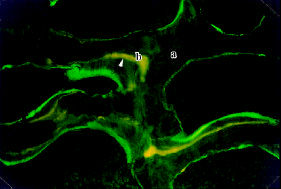
Tc2 group Treatment with Tc2 for 90 d had the
Fig 1. Trabecular bone of Tc2 group under fluorescent mi-
same effect on TA as Tc1, but for dynamic data,Tc2
croscope ( H-E stain, ×200). No yellow labeling was investi-
decreased LP and BFR and increased MAR, compared
gated in trabecular bone (a) except Tc high dose mg/kg for
with Tc1 group. The osteoclast number was signifi-
bone marker (b).
cantly decreased (-61 %) in Tc2 rats. No yellow label-ing was investigated in this dose ( 4.8 mg/kg) except
were atrophy. It was noted that the size of the uteri
high dose for bone marker under epifluorescent micro-
was significantly small and the endometrium was rela-
scope (Fig 1).
tively thiner in which there were fewer endometrial
Pathological changes (Fig 2)
glands and stroma.
Ova group The mild hydropic degeneration was
Estrone group The uteri in estrone group were
encountered in the liver cells and the epithelial cells of
increased in size and weight vs Ova group, the hydro-
the proximal convoluted tubules of the kidney. The uteri
pic degeneration was observed in the liver cells and the
Li QN et al / Acta Pharmacol Sin 2003 Jun; 24 (6): 599-604
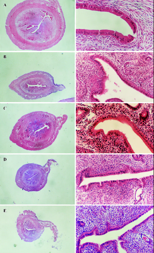
Fig 2. Cross-section of uterus and endometrium. Endometrium in sham (A) and estrone (C) was much thicker than that in
Ova (B), Tc1 (D), and Tc2 (E).
Li QN et al / Acta Pharmacol Sin 2003 Jun; 24 (6): 599-604
epithelial cells of the tubules of the kidney. The normal
Tc 30 mg⋅kg-1⋅d-1 is used mostly in a method to be
endometrium of uterine had been totally replaced by
labeled as a bone marker. If Tc is used in a dose lower
stratified squamous cells in 4 cases out of 9 (squamous
than 30 mg⋅kg-1⋅d-1 it will not have a yellow marker or
the marker will not be clearly seen on bone under
Tc2 group The hydropic degeneration was seen
epifluorescent microscope in early studies[4,5]. The lower
in the liver cells and the epithelial cells of the tubules of
doses of Tc in this study are 5 %-16 % dose of Tc as a
the kidney. Very mild fatty degeneration in the cells of
bone marker and 0.24 %-0.72 % dose of Tc as an anti-
liver was seen in 4 cases out of 9. The changes of uteri
biotic in clinic, which means this lower dose of Tc is
in Tc2 group were as the same as those in Ova group.
impossible to make bone and teeth yellow in adult rats,but for young rats is not clear. The data of lower doses
of Tc did not affect the labeling in higher dose as a bonemarker (Fig 1). Because the doses of Tc in this study
Tc in this study was found to be effective like
are very low, our hypothesis is that Tc is impossible to
estrogen on increasing trabecular bone mass in Ova rats.
cause serious side effects on liver and kidney. The re-
But the mechanisms between Tc and estrogen to pre-
sults of pathological section of the liver and kidney have
vent bone loss were different. Tc is a substance that
been verified. The most advantage of these doses of Tc
binds to calcium in bone surface and the mechanism of
is that it does not cause the proliferation of the uteri
Tc preventing bone loss is to decrease bone resorption,
(Fig 2). It is noted that the sizes of the uteri are signifi-
and in the meantime maintains bone formation indices
cantly small and the endometrium is relatively thiner in
in Ova rats. The effects of Tc are similar to anabolic
which there are fewer endometrial glands and stroma,
medicine, such as parathyroid hormone[9]. However,
while estrogen does enlarge uteri sizes due to their pro-
estrogen is a typical antiresorptive drug to decrease bone
liferation which may induce cancer of the uteri.
turnover, not only decreasing bone resorption but also
The results provide basic information about the
decreasing bone formation.
low doses and long-term effects of Tc on bone mass
The dynamic data indicated that Tc1 maintained
and bone structure, and can be used in designing the
bone formation indices almost at Ova level, such as LP,
future experiments. Tc structure can be modified to
which represents osteoblast recruitment. This result is
benefit the bone without severe side-effects on bone,
the same as that[2]. But MAR, which represents osteo-
teeth, liver, and kidney, etc[11].
blast activity has significantly been decreased in Tc1
In summary, this study indicated that two doses
compared with Ova group, and it is significantly lower
of hydrochloride tetracycline had the effects on pre-
than that in the estrogen group. Final result of Tc1 is to
vention of bone loss as well as showed no harms to
increase bone formation rate and reduce bone loss in
liver, kidneys, and uteri in ovariectomized rats. The study
Ova rats. Tc2 has the same effects on static data (TA,
showed an obvious advantage of Tc than estrogen,
TN, and TS) compared with Tc1, but dynamic data are
which induces uterus weight even it does prevent the
different from Tc1. Tc2 decreases LP and BFR/BV,
bone loss. This is the result of fundamental differences
but increases MAR. This occurrence shows that Tc2
of the mechanism in prevention of bone loss between
increases the osteoblast activity, while Tc1 increases
Tc and estrogen. As Tc does not slow down the bone
osteoblast recruitment. Why Tc1 and Tc2 have the dif-
formation, it indicates that low dosage of hydrochlo-
ferent bone activity on osteoblast is an interesting topic,
ride tetracycline may be a good medicine in the preven-
which needs further study. For the bone mass Tc2 al-
tion of bone loss in osteopenia induced by ovariectomy.
most has the same effects as Tc1. This result is prob-ably due to the dose differences of Tc being too small.
ACKNOWLEDGMENTS The authors are grateful to
The further study is neccessary to test more dose ef-
Prof FU Zhi-Gang (Department of Foreign Language
fects of Tc on bone. However, osteoclast numbers of
Teaching, Guangdong Medical College) for his excel-
both groups are significantly decreased in Ova rats. Tc2
lent English editorial assistance, and Prof CHEN Xiao-
had more suppressive effects on osteoclast than that in
Yi (Department of Pathology, Guangdong Medical
Tc1 group. The mechanism of Tc inhibiting osteoclast
College) for her investigation in soft tissue.
might be associated with its induction of apoptosis inosteoclast[10].
Li QN et al / Acta Pharmacol Sin 2003 Jun; 24 (6): 599-604
Jee WSS, Inoue J, Jee KW, Haba T. Histomorphometric as-
say of the growing long bone. In: Takahashi H, editor. Hand-
Li QN, Jee WSS, Ma YF, Ke HZ, Xie H, Huang LF, et al.
book of bone morphology. Japan: Niigata city; 1983. p 101-22.
Risedronate pretreatment does not hamper the anabolic ef-
Li QN, Liang NC, Huang LF, Wu T, Hu B, Mo LR. Skeletal
fects of prostaglandin E2 in OVX rats. Bone (Suppl) 1995;
effcts of constant and terminated use of sodium risedronate
17: 261s-6s.
in ovariectomized rats. Acta Pharmacol Sin 1998; 19: 160-3.
Sun TC, Mori S, Roper J, Brown C, Hooser T, Burr DB.
Li QN, Liang NC, Huang LF, Wu T, Hu B, Mo LE. Skeletal
Do different fluorochrome labels give equivalent histomorpho-
effects of constant and terminated use of risedronate on cor-
metric information? Bone 1992; 13: 443-6.
tical bone in ovx rats. J Bone Miner Metab 1999; 17: 18-22.
Lin BY, Jee WSS, Ma YF, Ke HZ, Kimmel DB, Li XJ.
Wronski TJ, Yen CF, Qi H, Dann LM. Parathyroid hormone
Effects of prostaglandin E2 and risedronate administration
is more effective than estrogen or bisphosphonates for res-
on cancellous bone in older female rats. Bone 1994; 15: 489-
Endocrinology 1993; 132: 823-30.
Hattner RS, Ilnicki LP, Hodge HC. The dose-response rela-
tionship of tetracycline to the detectability of labeled osteons
Grabowski PS. Tetracyclines induce apoptosis in osteoclasts.
by fluorescence microscopy. New York: deGruyter; 1977.
Bone 2000; 27: 75-80.
11 Sasaki T, Ohyori N, Debari K, Ramamurthy NS, Golub LM.
Parfitt AM, Drezner MK, Glorieux FH, Kanis JA. Bone
Effects of chemically modified tetracycline, CMT-8, on bone
histomorphometry nomenclature, symbols and units. J Bone
loss and structure and function in osteoporotic states. Ann N
Min Res 1987; 2: 596-610.
Y Acad Sci 1999; 878: 347-60.
Source: http://www.chinaphar.com/1671-4083/24/599.pdf
Computational Repositioning of the AnticonvulsantTopiramate for Inflammatory Bowel Disease Joel T. Dudley,1,2,3* Marina Sirota,1,2,3* Mohan Shenoy,4 Reetesh K. Pai,5Silke Roedder,1,3 Annie P. Chiang,1,2,3 Alex A. Morgan,1,2,3 Minnie M. Sarwal,1,3Pankaj Jay Pasricha,4 Atul J. Butte1,3† Inflammatory bowel disease (IBD) is a chronic inflammatory disorder of the gastrointestinal tract for which thereare few safe and effective therapeutic options for long-term treatment and disease maintenance. Here, we ap-plied a computational approach to discover new drug therapies for IBD in silico, using publicly available moleculardata reporting gene expression in IBD samples and 164 small-molecule drug compounds. Among the topcompounds predicted to be therapeutic for IBD by our approach were prednisolone, a corticosteroid used to treatIBD, and topiramate, an anticonvulsant drug not previously described to have efficacy for IBD or any related dis-
Prisoner Diabetes Handbook A Guide to Managing Diabetes— for Prisoners, by Prisoners published by the southern poverty law center Why A Handbook for Prisoners With Diabetes?Diabetes is important. It is common, chronic, and can cause disabling complications.What you do for yourself to take care of your diabetes is the most important factor in your diabetes being well controlled. Very little diabetes education is provided in prisons. There are few organized programs for prisoners with diabetes.Experience has shown that others with diabetes are a good source

