Checkpoint kinase 1-mediated phosphorylation of cdc25c and bad proteins are involved in antitumor effects of loratadine-induced g2/m phase cell-cycle arrest and apoptosis
MOLECULAR CARCINOGENESIS 45:461–478 (2006)
Checkpoint Kinase 1-Mediated Phosphorylationof Cdc25C and Bad Proteins Are Involved inAntitumor Effects of Loratadine-Induced G2/MPhase Cell-Cycle Arrest and Apoptosis
Jinn-Shiun Chen,1 Shyr-Yi Lin,2 Wei-Ling Tso,3 Geng-Chang Yeh,4 Wen-Sen Lee,5 How Tseng,6Li-Ching Chen,7 and Yuan-Soon Ho3*
1Division of Colon and Rectal Surgery, Department of Surgery, Chang Gung Memorial Hospital, Linkou, Taiwan2Department of Internal Medicine, School of Medicine, Taipei Medical University, Taipei, Taiwan3Graduate Institute of Biomedical Technology, Taipei Medical University, Taipei, Taiwan4Graduate Institute of Medical Sciences, and Department of Pediatrics, School of Medicine, Taipei Medical University,Taipei, Taiwan5Graduate Institute of Medical Sciences, and Department of Physiology, School of Medicine, Taipei Medical University,Taipei, Taiwan6Graduate Institute of Medical Sciences, School of Medicine, Taipei Medical University, Taipei, Taiwan7Institute of Biomedical Sciences, Academia Sinica, Taipei, Taiwan
In this study, we first demonstrated that loratadine (LOR), a promising world widely used oral anti-histamine,
effectively inhibits growth of tumors derived from human colon cancer cells (COLO 205) in an in vivo setting. In vitrostudy demonstrated that the anti-tumor effects of LOR in COLO 205 cells were mediated by causing G2/M phase cellgrowth cycle arrest and caspase 9-mediated apoptosis. Cell-cycle arrest induced by LOR (75 mM, 24 h) was associatedwith a significant decrease in protein levels of cyclin B1, cell division cycle (Cdc) 25B, and Cdc25C, leading toaccumulation of Tyr-15-phosphorylated Cdc2 (inactive form). Interestingly, LOR (75 mM, for 4 h) treatment alsoresulted in a rapid and sustained phosphorylation of Cdc25C at Ser-216, leading to its translocation from the nucleusto the cytoplasm because of increased binding with 14-3-3. We further demonstrated that the LOR-induced Cdc25C(Ser-216) phosphorylation was blocked in the presence of checkpoint kinase 1 (Chk1) specific inhibitor (SB-218078).
The cells treated with LOR in the presence of Chk1 specific inhibitor (SB-218078) were then released from G2/M arrestinto apoptosis. These results implied that Chk1-mediated phosphorylation of Cdc25C plays a major role in response toLOR-mediated G2/M arrest. Although the Chk1-mediated cell growth arrest in response to DNA damage is welldocumented, our results presented in this study was the first report to describe the Chk1-mediated G2/M cell-cyclearrest by the histamine H1 antagonist, LOR. ß 2006 Wiley-Liss, Inc.
Key words: loratadine; G2/M arrest; apoptosis; anti-tumor; Chk1
Cdc25C from ctyoplasm into the nucleus, and
The cell cycle is controlled by the periodic
checkpoint kinase 1 (Chk1), which phosphorylate
regulation of the highly conserved cyclin dependent
Cdc25C at Ser-216 and creates a consensus-binding
kinases (CDKs) [1]. In eukaryotes, the G
site for 14-3-3 [8]. The binding of 14-3-3 requires the
point is controlled by the cell-division cycle (Cdc)2/Cyclin B complex, whose activity is required forentry into mitosis [2]. Previous studies indicate thatCdc2 (Tyr-15) phosphorylation is maintained during
Abbreviations: Cdc 25B, cell-division cycle 25B; Chk1, checkpoint
cancer therapeutic drug-induced G
kinase 1; LOR, loratadine; ATR, ataxia telangiectasia-mutated and
Rad 3-related; FCS, Fetal calf serum; DBH, debromohymenialdisine;
mammalian cells [3–5]. Phosphorylation of Cdc2
DMSO, dimethylsulfoxide; PBS, phosphate-buffered saline; P.I.,
(Tyr-15) is accomplished by two major regulators
propidium iodide; FACS, fluorescence activated cell sorting; pNA,
including Wee1, which phosphorylate Cdc2 at Tyr-
p-nitroaniline; CCCP, carbonyl cyanide m-chlorophenylhydrazone;Val, valinomycin; I.P., intraperitoneal; TF, terfenadine; ATM, ataxia
15 and Myt1, which phosphorylate Cdc2 at Thr-14
and, to a lesser extent, Tyr-15 [6]. The Cdc2 (Tyr-15)
*Correspondence to: Graduate Institute of Biomedical Technol-
was activated by Cdc25C, a dual specific phospha-
ogy, Taipei Medical University, 250 Wu-Hsing Street, Taipei 110,Taiwan.
tase, whose activity is essential for entry into mitosis
Received 20 June 2005; Revised 22 September 2005; Accepted
[7]. In addition, two additional regulators include 14-
3-3, which binds Cdc25C and inhibits the import of
DOI 10.1002/mc.20165
ß 2006 WILEY-LISS, INC.
phosphorylation of Cdc25C at Ser-216, and mutat-
calf serum (FCS), penicillin/streptomycin solution,
ing this residue to Ala abolishes the interaction [9].
and fungizone were purchased from Gibco-Life
This site is present in the potential recognition motif
Technologies (Paisley, UK). The chemical inhibitors
for 14-3-3 and is phosphorylated in vitro by check-
were obtained from various sources as indicated: The
point kinases, such as Chk1 and Chk2 [10,11]. These
Chk1 inhibitors SB-218078 [21], and the Chk1/Chk2
results leads to the nuclear export of Cdc25C and
inhibitor debromohymenialdisine (DBH) [22,23]
its subsequent cytoplasmic sequestration by 14-3-3
were from Calbiochem (San Diego, CA).
protein, which prevents the activation of the down-stream target of Cdc25C, the cyclin B/Cdc2 kinase
that is responsible for G2/M transition. Thus, the
The following monoclonal antibodies were obtain-
association of 14-3-3 with target proteins could
ed from various sources as indicated: anti-caspase-8,
modulate cell-cycle progression through different
anti-cytochrome C, anti-cyclin B1, anti-Chk1 (sc-
mechanisms such as subcellular localization and
8408 or sc-7898), anti-Chk2 (sc-5278), anti-Cdc25B,
enzyme activity, depending on cellular signaling.
anti-GAPDH, anti-AIF, and anti-14-3-3 antibodies
It has been well demonstrated that Chk1 inhibi-
(Santa Cruz Biotechnology, Santa Cruz, CA), anti-
tion potentiate the cytotoxicity of DNA-damaging
phosphor BAD (Ser-155) (Cell Signaling Technolo-
drugs through abrogation of the cell-cycle check-
gy, Beverly, MA), anti-PCNA (Dako Corporation,
point [12–14]. However, several agents that including
Denmark), anti-caspase 9, anti-caspase 3 antibodies
anti-mitotic agent (paclitaxel) [15], topoisomerase II
(Stressgen Biotechnologies, Victoria, BC, Canada),
inhibitor (etoposide, camptothecin, and doxorubi-
anti-cytochrome C oxidase, (Research Diagnostics,
cin) [12,16], and microtubule-targeting agents
Flanders, NJ), and anti-b-actin (Sigma Aldrich Che-
(nocodazole) [17] which were not targeted on DNA
mie GmbH). The rabbit polyclonal antibodies
damage also revealed that G2/M arrest is largely
against phospho-Chk1 (Ser-345), phospho-Chk2
dependent on Chk1-mediated signaling pathway. In
(Thr-68), phospho-Cdc2 (Tyr-15), Cdc2, phospho-
this study, the Chk1 was demonstrated to play some
Cdc25C (Ser-216), and Cdc25C (C-20) were pur-
important role in the loratadine (LOR)-induced
chased from Cell Signaling Technology.
mitotic checkpoint. Our study also implied that theclinical efficacy of anti-tumorigenesis was expected
Cells and Culture Conditions
to be enhanced by Chk1 inhibitor, as evidenced byreleased of the G
The COLO 205 cell line was isolated from human
2/M phase arrest cells into apoptosis.
In the present study, we demonstrate that LOR
colon adenocarcinoma (American Type Culture
treatment causes a G
Collection CCL-222). The cell line FCH, a homo-
2/M phase cell-cycle arrest in
COLO 205 cells which is associated with a marked
zygous familial hypercholesterolemia cell (CRL 1831;
decrease in the expression of key G
American Type Culture Collection), was derived
proteins, including cyclin B1, Cdc25B, and Cdc25C.
from primary cultures of normal CRLs [24]. The cells
In addition, we provide evidence to indicate that
were grown in RPMI 1640 supplemented with 10%
cell-cycle arrest in LOR-treated COLO 205 cells is
FCS, penicillin (100 U/ml), streptomycin (100 mg/
caused by ataxia-telangiectasia-mutated and Rad3-
ml), and 0.3 mg/ml of glutamine for COLO 205;
related (ATR) /Chk1-mediated phosphorylation of
grown in DMEM/Ham's Nutrient Mixture F-12, 1:1
Cdc25C at Ser-216. Phosphorylation of Cdc25C in
with 2.5 mM L-glutamine, 1.2 g/L sodium bicarbo-
LOR-treated cells leads to its sequestration in the
nate, 15 mM HEPES and 0.5 mM sodium pyruvate
cytosol through increased binding with 14-3-3.
supplemented with 10% FCS, 10 ng/ml of cholera
Recent studies demonstrated that ATR/Chk1 is
toxin, 0.005 mg/ml of insulin, 0.005 mg/ml of
postulated in response to DNA damage by ionizing
transferrin, 100 ng/ml of hydrocortisone, and 10 mM
radiation, UV light, or interference with DNA
HEPES for CRL 1831 in a humidified incubator (378C,
replication [18–20]. In this study, our results provide
5% CO2). LOR (Sigma Chemical Co., St. Louis, MO)
further evidence showing that Chk1 signaling
was added at the indicated doses in 0.05% dimethyl-
regulatory proteins may be predominantly affect
sulfoxide (DMSO). For control treated cells, the same
the LOR-mediated G
volume of DMSO was added in a final concentration
cell-cycle checkpoint
of 0.05% (v/v) without LOR.
MATERIALS AND METHODS
Determination of Cell Viability
COLO 205 and CRL 1831 cells were treated with
Chemicals and Reagents
LOR (10–75 mM). Cell viability was determined at the
Protease inhibitors (phenylmethyl sulfonyl fluor-
indicated times based on 3-(4,5-dimethylthiazol-2-
ide (PMSF), pepstatin A, leupeptin, and aprotinin)
were acquired from Sigma Chemical Company
assay. Briefly, cells were seeded in a 96-well plate at
(Sigma Aldrich Chemie GmbH, Steinheim, Germany).
a density of 1 104 cells/well and allowed to adhere
Dulbecco's modified Eagle's medium (DMEM), Fetal
overnight. After removing the medium, 200 ml of
ANTITUMOR EFFECTS OF LOR IN HUMAN COLON CANCER CELLS GROWTH
fresh medium per well, containing 10 mmol/L HEPES
for translocation of cytochrome C from the mito-
(pH 7.4) was then added. Then, 50 ml of MTT was
chondria membrane into cytosol. Lysis of cells for
added to the wells and the plate was incubated for
mitochondrial protein extraction was performed in
2–4 h at 378C in the dark. The medium was removed
isotonic buffer (200 mM mannitol, 70 mM sucrose, 1
and 200 ml DMSO and 25 ml Sorensens's glycine buffer
mM EGTA, 10 mM HEPES, pH 6.9) by dounce
was added to the wells. Absorbance was measured
homogenization. Unbroken cells, nuclei, and heavy
using an ELISA plate reader at 570 nm.
membranes were pelleted at 1000g for 5 min anddiscarded. The mitochondrial enriched fraction was
Cell synchronization, drug treatment, and
collected by pelleting at 12000g for 20 min. The
flowcytometric analysis
pellet was then washed briefly in alkaline wash buffer
At 24 h after plating of cells, cells were washed
(0.1 M Na2CO3, pH 11.5) to separate peripherally
twice with phosphate-buffered saline (PBS) and then
associated (alkali sensitive) mitochondrial proteins
incubated with medium containing 0.04% FCS for
from membrane integrated (alkali resistant) mito-
additional 24 h. Under such conditions, cells were
chondrial proteins and centrifuged. The pellet (con-
arrested in G0/G1 as determined by using flowcyto-
taining the membrane integrated proteins) was
metric analysis [25]. After serum starvation, the low-
resuspensed in RIPA lysis buffer (1 PBS, 1% Nonidet
serum (0.04% FCS) medium was removed and the
P-40, 0.5% sodium deoxycholate, 0.1% sodium
cells were then challenged by addition of medium
dodecyl sulphate) with protease inhibitor cocktail
containing 10% FCS. LOR solutions were prepared in
(Calbiochem) and used for immunoblotting. For
a final concentration of 0.05% (v/v) DMSO. The cell-
cytochrome C oxidase protein detection (cyto-
cycle stages in the LOR and DMSO-treated groups
chrome C oxidase, subunit IV, detection was
were measured by flowcytometric analysis. Cells
employed as a control to demonstrate that mito-
were harvested and stained with propidium iodide
chondrial protein fractionation was successfully
(P.I.) (50 mg/ml) (Sigma Chemical Co.), and DNA
isolated), equivalent samples (20 ml containing
content was measured using a fluorescence activated
approximately 50 mg protein) were separated by
cell sorting (FACS) can laser flowcytometry analysis
SDS–PAGE on 12% Tris glycine gels and transferred
system (Becton–Dickinson, San Jose, CA); and 15000
to 0.2 mM polyvinylidene difluoride membranes
events were analyzed for each sample.
(Invitrogen). Blots were probed with a mouse mono-clonal antiserum specific for cytochrome C (Santa
Western Blotting Analysis
Cruz, CA) or with a rabbit polyclonal antibody specific
Western blotting analysis was performed as
for cytochrome C oxidase followed by the appropriate
described previously [26]. Briefly, cell lysates were
secondary antibodies conjugated to horseradish per-
prepared, electrotransferred, immunoblotted with
oxidase (Santa Cruz) and then visualized by Super-
antibodies, and then visualized by incubating with
Signal chemiluminescence kit as described in the
the colorigenic substrates (nitroblue tetrazolium,
manufacturer's protocol (Pierce Biotechnology, Rock-
NBT and 5-bromo-4-chloro-3-indolyl phosphate
ford, IL) and visualized by autoradiography.
(BCIP)) (Sigma Chemical Co.). The expression of
Preparation of Nuclear and Cytoplasmic Fractions
either GAPDH, b-actin, or cytochrome C oxidase wasused as control for equal protein loading.
Nuclear and cytoplasmic fractions from control
(DMSO-treated) and LOR-treated COLO 205 cellswere prepared as described previously [5]. Briefly,
cells were harvested by scraping and rinsed twice in
Equal amounts of protein were immunoprecipi-
ice-cold PBS. The cells were then swollen in ice-cold
tated with saturating amounts of anti-14-3-3 anti-
hypotonic lysis buffer (20 mM HEPES, pH 7.1, 5 mM
body. Immunoprecipitates were washed five times
KCl, 1 mM MgCl2, 10 mM N-ethylmaleimide, 0.5 mM
with extraction buffer and once with PBS. The pellet
phenylmethylsulfonyl fluoride, 5 mg/ml of pepstatin
was then resuspended in sample buffer (50 mM Tris,
A, 2 mg/ml of chymostatin, 5 mg/ml of leupeptin,
pH 6.8; 100 mM dithiothreitol; 2% SDS; 0.1%
5 mg/ml of aprotinin, 5 mg/ml of anti-pain) for 10 min.
bromophenol blue; 10% glycerol) and incubated
The cells were lysed by 20 strokes in a Dounce
for 10 min at 908C before electrophoresis to release
homogenizer, and the nuclei were cleared by centri-
the proteins from the beads. The 14-3-3-immuno-
fugation (400g, 10 min). After this step, the super-
precipitated phosphor Cdc25C and phosphor BAD
natant (cytosolic fraction) was concentrated and
proteins were then measured by Western blot
stored at 808C. The nuclear extract was prepared
analysis using specific antibodies.
using the same lysis buffer and stored at 808C priorto Western blot analysis for Cdc25C. The blot was
Isolation of Mitochondria and Cytosolic
stripped and reprobed with b-actin or PCNA antibody
Fractions of Cell Lysates [27]
to ensure equal protein loading as well as to rule out
The COLO 205 cells were incubated with LOR (25
cross contamination of cytoplasmic and nuclear
mM) for the indicated time points and then assayed
Chk1 Kinase Activity Assay
the lysis buffer and once with kinase buffer contain-
Approximately 106 cells were plated at a conflu-
ing 50 mM Tris-HCl (pH 7.4), 10 mM MgCl2, and
ence of 70% and exposed to LOR (75 mM, 4 h). Cells
1 mM dithiothreitol. The beads were incubated with
were collected, and Chk1 kinase activity was mea-
50 ml of kinase reaction mixture containing 50 mM
sured by a immunoprecipitation kinase assay as
Tris-HCl (pH 7.4), 10 mM MgCl2, 1 mM dithiothrei-
described previously [28,29]. Briefly, the Chk1 assay
tol, 10 mM ATP, 5 mCi of [g-32p] ATP, and 0.5 mg/ml
was carried out in 50 ml volume in the following
of histone H1 for 15 min. The reaction was
buffer: 20 mM Na-HEPES, pH 7.4, 50 mM KCl, 1 mM
terminated by the addition of 20 ml of 4 Laemmlis
sample buffer and boiling for 5 min. The 32p-
2, and 1 mM DTT. A synthetic
peptide with a sequence flanking Ser-216 of human
phosphorylated histone H1 was separated by 0.1%
Cdc25C, KVSRSGLYRSPSMPENLNRK, was used as its
SDS, 10% polyacrylamide gel, and determined by
substrate [29]. Chk1 was used at 5 nM, the peptide
autoradiography using Kodak X-Omat film.
was used at 20 mM, and [g-32P]ATP was used at 10 mM(final concentration). Reactions were incubated at
Analysis of Apoptosis
378C for 20 min and stopped by adding 5 ml of acetic
Apoptosis in the COLO 205 cells subjected to
acid containing 10 mM ATP. Thirty-microliter
various treatments was determined by using the
samples were spotted on 2-cm phosphocellulose
DNA fragmentation analysis [33]. Briefly, the LOR
filter disks, which were washed four times, 5 min
and DMSO-treated cells were seeded on 100-mm
each with 0.5% phosphoric acid and counted by
dishes. The DNA was extracted twice with equal
liquid scintillation. The apparent Km for ATP in the
volumes of phenol and once with chloroform-
Chk1 assay was about 120 mM.
isoamyl alcohol (24:1 v:v), precipitated with 0.1volume of sodium acetate, pH 4.8, and 2.5 volumes
Wee1 Kinase Activity Assay
of ethanol at 208C overnight, and finally centri-
The recombinant Cdc2 protein was purified as a
fuged at 13000g for 1 h. Genomic DNA was
glutathione S transferase fusion protein containing
quantitated, and equal amounts of DNA sample in
full-length human Cdc2 (Santa Cruz Biotechnology)
each lane were electrophoresed in a 2% agarose gel.
and then used as a substrate for Wee1 kinase assay.
The DNA was visualized by ethidium bromide
Glutathione S-transferase protein was used as a
negative control substrate and was prepared accord-ing to the standard procedure suggested by the
Caspase Activity Assay
manufacturer (Pharmacia, Piscataway, NJ). Prepara-
Caspase activity was measured by using caspases 3
tion of cell lysates, immunoblotting, immunopreci-
(Promega, Madison, WI) and 9 (Chemicon, Teme-
pitation were performed as described previously [25]
cula, CA) colorimetric activity assay kits as previous
except that cell lysis buffer for immunoprecipitation
described [34,35]. Briefly, COLO 205 cells were lysed
was 50 mM Tris-HCL, pH 7.5; 1% Triton X-100;
by addition of cell lysis buffer and protein concen-
0.5 mM Na3VO4; 50 mM sodium fluoride; 5 mM
tration was measured. Caspase activity was assayed at
sodium pyrophosphate; 10 mM sodium 2-glycero-
378C in 100 ml of assay buffer containing 50 mg (for
phosphate; 0.1 mM PMSF; 1 mg/ml of aprotinin; 1 mg/
caspase 3) or 30 mg (for caspase 9) of the indicated
ml of pepstatin; 1 mg/ml of leupeptin; 1 mM micro-
colorimetric peptide. Caspase activity was measured
cystin; 1 mM DTT; 1 mM EDTA, and 1 mM EGTA.
by the release of p-nitroaniline (pNA) from the
Kinase assays for Wee1 were performed as described
labeled substrates Ac-DEVD-pNA and Ac-LEHD-
previously [30,31]. The Wee1 kinase assay used 500 mg
pNA for caspase 3 and 9, respectively, and the free
of cell extract for immunoprecipitating kinase, and
pNA was quantified at 405 nm.
2 mg of relative protein substrate. Kinase reaction wascarried out by incubation for 30 min at 308C and
Mitochondrial Transmembrane Potential Assay
terminated by addition of 30 ml of 6 Laemmli SDS
To assess the mitochondrial transmembrane
sample buffer [32]. Substrate phosphorylation was
potential (DCm), COLO 205 cells (1 106) were
analyzed by SDS-polyacrylamide gel electrophoresis
seeded in a six-well plate washed twice with PBS
and then loaded with the cationic lipophilic fluor-ochrome JC-1 (5 mg/ml) for 10 min at 378C. Cells were
Cdc2 Kinase Activity Assay
washed twice with PBS and submitted to FACS-
The method for the assay of the Cdc2 kinase was
analysis. The red fluorescence of JC-1 aggregates
according to our previous report [26]. The protein
corresponds to the mitochondrial membrane poten-
content in each sample was determined as described
tial whereas the green fluorescence of JC-1 mono-
above and adjusted to 100 mg/lane. Lysate (in 0.5 ml
mers is indicative for the mitochondrial mass. Active
of extraction buffer) was immunoprecipitated with
mitochondria with high DCm accumulate JC-1
5 mg monoclonal anti-cyclin B1 antibody, the
aggregates, which are red, whereas, in the mitochon-
immuno-complexes were washed three times with
dria with low DCm, JC-1 stays in a monomeric, green
ANTITUMOR EFFECTS OF LOR IN HUMAN COLON CANCER CELLS GROWTH
form. This renders the red/green ratio, a sensitive
indicator of the mitochondrial DCm changes. Inaddition, carbonyl cyanide m-chlorophenylhydra-
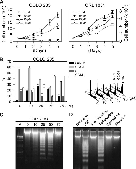
To investigate the antiproliferative effects of LOR,
zone (CCCP; Calbiochem) or valinomycin (Val,
human colon cancer cells (COLO 205) and human
Sigma) were dissolved in 100% acetone and diluted
normal CRL 1831 were treated with different doses
in complete medium; the acetone concentration in
(10–50 mM) of LOR for 1–5 days and the cell growth
the medium did not exceed 1%. Both of the CCCP
numbers were then determined (Figure 1A). LOR at
and Val were added at a final concentration of 200 mM
lower dose (10 mM) suppressed COLO 205 cell
as a positive control, and the fluorescence was
proliferation while at higher dose (25 mM) induced
assessed for each time point, a red/green fluorescence
cell death (Figure 1A, left). Interestingly, such effect
ratio was then calculated. The mean red fluorescence
was less profound when normal human colon
of drug-treated cells was measured at 0, 2, 4, 6, 8, 10,
epithelial cells (CRL 1831) treated with LOR (Figure
and 12 h after LOR treatment, and presented as a
1A, right). These results indicated that colon cancer
ratio of the absorbance in 590/535 nm [36].
cells were more susceptible to LOR treatment thannormal human CRLs.
Immunofluorescence Staining And
To further demonstrate whether the cytotoxic
Confocal Microscopic Observation
effect induced by LOR was due to apoptotic cell
For microscopic observations of nuclear/cyto-
death. The LOR-induced apoptotic effect was eval-
plasmic and nuclear/mitochondria distribution of
uated by flowcytometric and DNA fragmentation
Cdc25C and AIF, respectively, COLO 205 cells were
analysis. As shown in the Figure 1B, both of the sub-
incubated in 0.05% DMSO (control) or LOR for the
G1 and G2/M phase populations were observed in
indicated time points. COLO 205 cells were immu-
COLO 205 cells exposed to 20–50 mM of LOR.
nostained with monoclonal antibodies specific to
Interestingly, G2/M arrest instead of DNA fragmen-
phosphor Cdc25C (Ser-216) and AIF (Santa Cruz, CA)
tation was observed in COLO 205 cells, which
for 2 h. After washing with PBS/Tween 20 for three
exposed to higher dose (75 mM) LOR. Our previous
times, monoclonal antibodies were visualized with
report have demonstrated that apoptosis and G0/G1
goat-anti-mouse IgG antibody labeled with FITC
cell-cycle arrest was induced by terfenadine (TF), an
(green) for 1 h. Propidium iodide at concentration of
histamine H1 receptor antagonist with a mechan-
100 ng/ml for 5 min was used for nuclear staining.
isms similar to LOR [37,38]. In order to demonstrate
Stained cells were imaged by confocal scanning
whether the LOR- or TF-induced apoptosis was
microscopy (Olympus, Tokyo, Japan) using excita-
mediated through the blockage of the specific
tion/emission wavelengths of 458/488 nm and 543/
histamine H1 receptor signaling pathway, four
633 nm for FITC and P.I., respectively.
additional histamine H1 blockers including cetiri-zine, ebastine, epinastine, and fexofenadine [39]
Treatment of COLO 205-Derived Xenografts In Vivo
were added to COLO 205 cells (75 mM, 24 h) thendetected for DNA laddering effect. Our data demon-
COLO 205 cells were grown in RPMI 1640 supple-
strated that DNA fragmentation was observed only
mented with 10% FCS as described in our previous
in the LOR- and TF-treated group (Figure 1D). Such
studies [25–27]. Cells were harvested through two
results suggested that the histamine H1 receptor was
consecutive trypsinizations, centrifuged at 300g for
not the major apoptosis-signaling sensor in response
5 min, washed twice, and resuspended in sterile PBS.
to LOR in the COLO 205 cells.
Cells (5 106) in 0.2 ml were injected subcuta-
To address the cell-cycle effects of LOR, the COLO
neously between the scapulae of each nude mouse
205 cells were synchronized at the G0/G1 phase by
(purchased from National Science Council Animal
0.04% serum starvation for 24 h [25,26]. After serum
Center, Taipei, Taiwan). After transplantation,
starvation, the complete medium containing 10%
tumor size was measured using calipers and the
FCS was then replaced. The cells treated either with
tumor volume was estimated according to the
mock-treated (DMSO) or LOR (75 mM) were then
formula tumor volume (mm3) ¼ L W2/2, where L
measured for cell cycle by flowcytometric analysis.
is the length and W is the width [26]. Once tumors
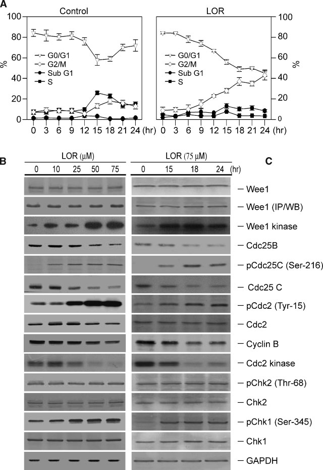
As shown in Figure 2A (right), G2/M cell-cycle arrest
reached a mean size of 200 mm3, animals received
was observed initially at 9 h and reached the
intraperitoneal (I.P.) injections of either 25 ml DMSO
maximal level of more than 40% at 24 h after LOR
or 25 mg/kg LOR three times per wk for 6 wk.
It is well established that onset of mitosis is
triggered by activation of the Cyclin B1/Cdc2 kinase,
Statistical analysis was carried out using analysis of
which is absolutely required for transition of G2 cells
variance (ANOVA)—one way analysis of variance
into M phase [19,40]. During the G2 phase, Cyclin
with Student—Newman correction, and the Stu-
B1/Cdc2 is inactivated by phosphorylation at the
dent's t-test. Significance was assumed for values of
Thr-14 and Tyr-15 residues, which are dephosphory-
P < 0.05.
lated by Cdc25C phosphatase before going into
Figure 1. Cell growth inhibitory effects of LOR on human colon
using established CellFIT DNA analysis software. Three samples were
cancer and normal epithelial cells. (A) Human colon cancer (COLO
analyzed in each group, and values were presented as mean SE. (C)
205) cells and human normal CRL 1831 were treated with various
COLO 205 cells were treated with LOR (10–75 mM) or DMSO
concentrations of LOR (10–50 mM) for 5 days. Media with or without
(0.05%), and DNA fragmentation assay performed 24 h later. M,
LOR were changed everyday until cell counting. Three samples were
molecular weight marker. (D) COLO 205 cells were cultured in media
analyzed in each group and the results were presented as
supplemented with 10% FCS in the presence of histamine H1
means SE. (B) Dose-dependent response of LOR on cell-cycle
receptor antagonists namely LOR, TF, cetirizine, ebastine, epinastine,
regulation. COLO 205 cells were cultured in media supplemented
and fexofenadine (75 mM each treated for 24 h). DNA fragmentation
with 10% FCS and LOR (10–75 mM) for 24 h. Percentage of cells in
analysis was then performed as described above.
sub-G1, G0/G1, S, and G2/M phases of the cell cycle were determined
mitosis. In this study, our results indicated that cells
[26,41] were selected as 0 h (representing the G0/G1
arrested at the G2/M phase by 24 h of LOR (10–75 mM)
phase), 15 h (representing the S phase), 18 h
treatment were regulated by inhibition of the
(representing the G2/M phase), and 24 h (represent-
cyclin B1/cdc2 protein levels and its kinase activity
ing the 2nd G0/G1 phase). The cell lysates were then
(Figure 2B). In order to examine the time-dependent
analyzed for expression of cell-cycle regulatory
effect of LOR on G2/M phase cell-cycle arrest, the
proteins by immunoblotting using specific antibo-
COLO 205 cells were synchronized at the G0/G1
dies (Figure 2C). To assess the Cdc2 kinase activity,
phase by 0.04% serum starvation for 24 h [25,26].
the level of phosphorylated Cdc2 (Tyr-15) as well as
After serum starvation, the complete medium con-
the level and/or kinase activity of the Wee1 and
taining 10% FCS was then replaced. The time points
Cdc25C phosphatase, two key enzymes that regulate
according to Figure 2A and our previous studies
the Cdc2 kinase activity, were determined in the
ANTITUMOR EFFECTS OF LOR IN HUMAN COLON CANCER CELLS GROWTH
Figure 2. Effects of LOR on cell-cycle regulations in human COLO
regulatory proteins in COLO 205 cells. The cells were rendered
205 cells. (A) Time-dependent effects of LOR in human COLO 205
quiescent by incubation for 24 h in the cultured media containing
cell-cycle analysis. FACS analysis of DNA content was conducted
0.04% FCS. After 24 h, cells were released from quiescence by
after COLO-205 cells release from quiescence by incubation in
incubation in culture media supplement with 10% FCS and 0.05%
culture media supplemented with 10% FCS in the presence of
DMSO with or without LOR (10–75 mM) for additional 24 h. The cells
0.05% DMSO or LOR (75 mM) at the indicated time points.
were harvested, lysed, and the levels of G2/M phase regulatory
Percentage of cells in different phases of cell cycle was determined
proteins and its kinase activities were determined as described in the
using established CellFIT DNA analysis software. Three samples were
Materials and Methods. Membrane was also probed with anti-
analyzed in each group, and values represent the mean SE. (B)
GADPH antibody to correct for difference in protein loading.
Dose-dependent effects of LOR on the expression of G2/M phase
LOR-treated COLO 205 cells. The results indicated
The Chk1 is a serine-threonine kinase that is
that phosphorylation of Cdc2 (Tyr-15) and its kinase
critical for G2/M arrest in response to DNA damage.
activity was changed as early as 15 h after LOR
Recently, it has been demonstrated that Chk1
(75 mM) treatment (Figure 2C). Consistent with the
inactivate the pro apoptotic protein BAD by phos-
effect on phosphorylation of Cdc2 (Tyr-15), LOR
phorylating residues critical for BAD functions
treatment also resulted in a marked induction of
in vitro [46]. In this study, our results demonstrated
Wee1 kinase activity in the same levels of total Wee1
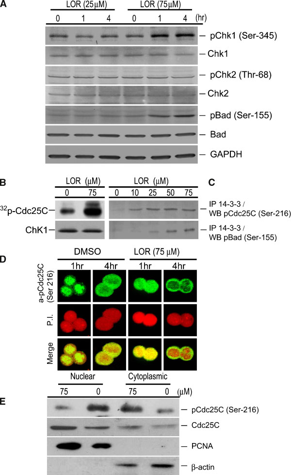
that BAD (Ser-155) was phosphorylated as early as 1 h
protein (Figure 2B and C). In addition, the LOR
in COLO 205 cells by LOR treatment (Figure 3A).
treatment in COLO 205 cells also resulted in a
Because phosphorylation of Cdc25C (Ser-216) [43]
significant reduction in Cdc25C protein levels
and BAD (Ser-155) [47] creates a binding site for 14-3-
(Figure 2B and C). Thus, the induction of Wee1
3, we then examined the effect of LOR on the binding
kinase at 15 h after LOR (75 mM) treatment in COLO
of Cdc25C and BAD with 14-3-3 (Figure 3C). The
205 cells was associated with the decreased of the
lysate proteins from control and LOR-treated cells
Cdc25C phosphatase activity which eventually
(10–75 mM for 4 h) were immunoprecipitated using
increased the level of phosphorylated Cdc2 (Tyr-15)
anti-14-3-3 antibody, and the immune complex was
(Figure 2C). The net effect of the results is a G2/M
analyzed for the presence of Cdc25C and BAD by
arrest following LOR treatment.
immunoblotting. As can be seen in Figure 3C, LOR
Chk1 and Chk2 are intermediaries of DNA damage
treatment resulted in increased binding of Cdc25C
checkpoints and activated by phosphorylation on
and BAD with 14-3-3 at the 4 h time point. These
Ser-345/Ser-317 and Thr-68, respectively [20,42].
results suggested that LOR treatment might lead to
The Chk1 and Chk2 kinases have been reported to
translocation of Cdc25C from the nucleus to the
inhibit Cdc25C kinase activity by phosphorylation at
cytoplasm because of increased binding with 14-3-3.
the Ser-216 residue [43–45]. We therefore examined
We examined this possibility by immunohistochem-
whether LOR treatment affects the phosphorylation
istry, and the data were shown in Figure 3D. Cells
of Chk1 or Chk2. Representative immunoblots for
were treated with DMSO (control) or 75 mM LOR for 4
phospho-Chk1 showed increased Ser-345 phosphor-
h, and then stained with anti-pCdc25C (Ser-216)
ylation of Chk1 at 15 h after LOR (75 mM) treatment,
antibody (FITC, green) or nucleic acid binding dye
whereas the level of total Chk1 proteins were not
P.I., red. In LOR (75 mM, 4 h)-treated cells, pCdc25C
affected (Figure 2C). The total and its phosphory-
(Ser-216) was localized in the cytoplasm (green
lated form of the Chk2 (Thr-68) proteins did not
staining surrounding P.I.-stained nuclei) as well as
changed in the COLO 205 cells even at 24 h after LOR
in the nucleus (red staining in nucleus). In contrast,
(75 mM) treatment (Figure 2B and C).
the nuclei of the control cells were stained with
To investigate the early stage responses of the
pCdc25C (Ser-216) (green), indicating translocation
COLO 205 cells for LOR treatment, the total and its
of pCdc25C (Ser-216) from the nucleus to the
phosphorylated form of the Chk1 (Ser-345), Chk2
cytoplasm (Figure 3D). Cytoplasmic accumulation
(Thr-68), and Bad (Ser-155) were then determined. As
of pCdc25C (Ser-216) upon treatment with LOR was
shown in the Figure 3A, our study demonstrated that
confirmed by biochemical fractionation of cytoplas-
Chk1 (Ser-345) but not Chk2 (Thr-68) was phos-
mic, and nuclear fractions from control (DMSO-
phorylated in the COLO 205 cells as early as 1 h when
treated) and LOR treated (75 mM for 4 h) cells
exposed to higher dose LOR (75 mM). The kinase
followed by immunoblotting using anti-pCdc25C
activity of Chk1 was then determined in the lysates
(Ser-216) antibody, and the results were shown in
prepared from DMSO- and LOR-treated cells (75 mM
Figure 3E. The total protein level of Cdc25C was also
for 4 h) using GST-Cdc25C as a substrate. As can be
determined in different fractions of the LOR- and
seen in the Figure 3B, the Chk1 kinase activity was
control-treated cells, which represented as a protein
significantly higher in LOR-treated cells than in
loading control. A 4 h time point was selected to
control cells.
minimize influence of LOR induced decline in
Figure 3. Effect of LOR on binding of Cdc25C and BAD with 14-3-
(10–75 mM for 4 h) were used for immunoprecipitation with anti-14-
3 and on nuclear/cytoplasmic distribution of Cdc25C. (A) Immuno-
3-3 antibody followed by Western blotting (WB) for phosphor
blotting assay for effect of LOR on protein level, and phosphorylation
Cdc25C (Ser-216) and phosphor BAD (Ser-155). (D) Confocal
of Chk1 (Ser-345) and BAD (Ser-155). COLO 205 cells were cultured
microscopic analysis for nuclear/cytoplasmic distribution of Cdc25C
in the presence of 75 mM LOR for the indicated time periods. The
(Ser 216) in control (DMSO-treated) and LOR-treated cells. Cells were
blots were stripped and reprobed with anti-GAPDH antibody to
treated with DMSO (control) or 75 mM LOR for 1 or 4 h and then
ensure equal protein loading. (B) Effect of LOR on Chk1 kinase
stained with anti-Cdc25C (Ser-216) antibody (green) or P.I. (red). (E)
activity. COLO cells were treated with DMSO or 75 mM LOR for 4 h.
Immunoblotting assay for pCdc25C(Ser-216) and Cdc25C using
Chk1 was immunoprecipitated from the lysates of control and LOR-
nuclear and cytoplasmic fractions prepared from control (DMSO-
treated cells, and the kinase activity was determined using
treated) and LOR-treated cells (75 mM for 4 h). Blots were stripped
synthesized Cdc25C peptide as a substrate described in Materials
and reprobed with anti-b-actin and anti-PCNA antibodies to
and Methods. The membrane was probed with anti-Chk1 antibody
normalize for equal protein loading as well as to rule out cross
to ensure equal protein loading. (C) Effect of LOR on binding of
contamination of the nuclear and cytoplasmic fractions. [Color figure
phosphor Cdc25C (Ser-216) and phosphor BAD (Ser-155) with 14-3-
can be viewed in the online issue, which is available at www.
3. The protein lysates (200 mg) from control and LOR-treated cells
ANTITUMOR EFFECTS OF LOR IN HUMAN COLON CANCER CELLS GROWTH
Cdc25C protein level shown in the Figure 2B. In
containing 4N-DNA (G2/M phase cells) (Figure 4B,
DMSO-treated control, the intensity of Cdc25C
immunoreactive band was significantly higher in
To further clarify the role of checkpoint kinases
the lane corresponding to nuclear fraction than in
activation on LOR-mediated G2/M arrest, two inhi-
the cytoplasmic fraction (Figure 3E). Treatment of
bitors were adapted in our experiment including
cells with LOR (75 mM) resulted in a decrease in
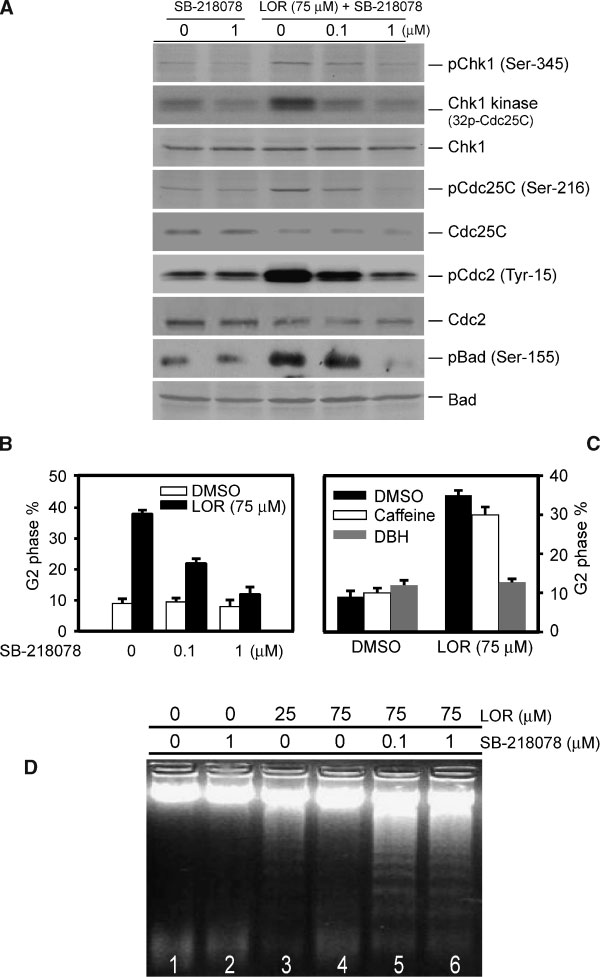
DBH [48] and caffeine [49–51], which inhibited
nuclear Cdc25C signal intensity with a concomitant
the Chk1/Chk2 and ataxia telangiectasia-mutated
increase in cytoplasmic Cdc25C signal intensity
(ATM)/ATR kinases, respectively. Interestingly, the
(Figure 3E). The blot was stripped and reprobed with
LOR-mediated G2/M arrest was completely attenu-
anti-b-actin and anti-PCNA antibodies to determine
ated by DBH (10 mM) (Figure 4C, gray bars), but not
cross contamination, if any, of the nuclear and
by caffeine (5 mM) (Figure 4C, open bars). Such
cytoplasmic fractions and to ensure equal protein
results implied that at least part of the LOR-induced
loading (Figure 3E). These results confirmed that
G2/M arrested cells utilize a caffeine-insensitive
LOR treatment, indeed, promoted translocation of
pathway in which checkpoint signaling by ATM or
Cdc25C from the nucleus to the cytoplasm.
ATR was most likely not involved.
To determine the possible role of Chk1 kinase
To examine the effect of Chk1 on LOR-mediated
activation in the regulation of LOR-mediated G2/M
apoptotic effects, COLO 205 cells were treated with
arrest, we examined the effects of Chk1 kinase on
LOR (25 and 75 mM) and then incubated for 24 h in
LOR-induced G2/M arrest in COLO 205 cells using
the medium containing 0.1–1 mM SB-218078 (Figure
Chk1-specific inhibitor SB-218078 [21]. We first
4D, lanes 5 and 6) or, as a control, 0.05% DMSO
demonstrated that SB-218078 (0.1 mM) inhibited
(Figure 4D, lane 1). The presence of DNA fragmenta-
the phosphorylation of Chk1(Ser-345) and its kinase
tion was detected as described above. The dose of SB-
activity in COLO 205 cells when treated with LOR
218078 used in this study (0.1–1 mM) was found to
(Figure 4A). The inhibitory effects of SB-218078 on
maximally prevent the LOR-induced G2/M arrest,
LOR-induced Cdc25C, Cdc2, and BAD phosphoryla-
which resulted in DNA laddering formation (Figure
tion were demonstrated (Figure 4A). To determine
4D, lanes 5 and 6). As a control, a parallel set of COLO
the effective dose of SB-218078 on LOR-mediated G2/
205 cells was incubated in the presence of 1 mM SB-
M arrest response, COLO 205 cells were treated with
218078, no significant DNA fragmentation was
LOR and then cultured for 24 h in the presence of
observed (Figure 4D, lane 2).
SB-218078 (0.1 and 1 mM). The resulting cells were
Our results revealed that lower dose (10–50 mM)
analyzed for DNA contents by FACS flowcytometric
LOR could induced both the G2/M arrest and
analysis as described in Materials and methods. The
apoptosis in COLO 205 cells (Figure 1B and D). The
results showed that the LOR-induced G2/M arrested
mechanisms of LOR-mediated apoptosis need to be
cells (Figure 4B, solid bar) was 3.9-fold higher than
further investigated. Since the mitochondria signal-
that in the control cells (Figure 4B, open bar).
ing regulatory proteins seems to be required for
Furthermore, incubation of LOR-treated COLO
different anti-cancer agents-induced apoptosis in
205 cells with SB-218078 resulted in a marked
COLO 205 cells [35,52–54]. We first examined
attenuation of LOR-induced G2/M arrest in a dose-
whether cytochrome C release from mitochondria
dependent manner (Figure 4B, solid bars). Incuba-
into the cytosol and dissipation of the electrochemi-
tion with 0.1 mM SB-218078 inhibited the LOR-
cal gradient (DCm) was involved in the LOR-
induced G2/M arrest by approximately 50%, while
mediated apoptosis. This was monitored by staining
incubation with 1 mM SB-218078 diminished the
the COLO 205 cells with JC-1, a fluorescent dye,
LOR-induced G2/M cell-cycle arrest by 80% (Figure 4B,
which differentially stains mitochondria in accor-
solid bars). No further inhibitory effect on LOR-
dance to their DCm. Active mitochondria with high
induced G2/M arrest was seen when higher doses of
DCm accumulate JC-1 aggregates, which are red
SB-218078 were used (data not shown). Incubation
(Figure 5A, 0 h), whereas, in the mitochondria with
of DMSO-treated COLO 205 cells with SB-218078
low DCm, JC-1 stays in a monomeric, green form
had no detectable effect on the population of cells
(Figure 5A, 12 h). This renders the red/green ratio, a
Figure 4. The Chk1 regulatory effects of LOR-induced G2/M cell
content, indicative of G2/M phase of the cell cycle, is shown as the
cycle arrest. (A) COLO 205 cells were treated with LOR (75 mM) in the
mean SE from three independent experiments. (C) COLO 205 cells
presence of SB-218078 (0.1 and 1 mM) for 24 h. The treated cells
were treated with LOR (75 mM) followed by incubation for 24 h in
were collected, lysed, and the level of phosphorylated Chk1 (Ser-
medium containing 0.05% DMSO (solid bars), 5 mM caffeine (open
345), Cdc25C (Ser-216), Cdc2 (Tyr-15), and BAD (Ser-155) as well as
bars), or 10 mM DBH (gray bars). Cell samples were analyzed for DNA
the level of total Chk1, Cdc25C, Cdc2, and BAD in each sample was
content by flowcytometry. The percentage of G2/M phase cells
determined by immunoblotting analysis with relevant specific
shown represents the average of three independent experiments. (D)
antibodies. The Chk1 kinase activity was determined as described
COLO 205 cells were incubated for 24 h in medium containing
in Materials and Methods. (B) Cells were treated with DMSO (open
0.05% DMSO, SB-218078 alone, or combine treatment with LOR
bars) or LOR (75 mM) (solid bars), and incubated for 24 h in the
and SB218078. The DNA samples were isolated from cells and
presence of Chk1 inhibitor (SB-218078). The resulting cells were
analyzed for DNA fragmentation as described in the Material and
harvested, stained with PI and analyzed for DNA contents by
flowcytometry as described above. Percentage of cells with 4N-DNA
ANTITUMOR EFFECTS OF LOR IN HUMAN COLON CANCER CELLS GROWTH
sensitive indicator of the mitochondrial DCm
ptotic responses by which the initiator (caspase 9
changes, which does not depend on other factors
and 8) and the effector (caspase 3) were determined
such as mitochondrial size, shape, and density,
by immunoblotting and caspase activity analysis
which may influence single-component fluorescent
(Figure 6A and B). The COLO 205 cells were treated
signals. Analyzed in a real-time plate reader assay,
with various concentrations of LOR (10–75 mM) for
DCm stayed relatively stable in untreated COLO 205
24 h. Our data demonstrated that LOR at a lower dose
cells, while it was rapidly (within 6 h) dissipated by
(25–50 mM) activated the caspase 3 as evidenced
25 mM LOR treatment (Figure 5A, lower panel). The
by a decreased in the protein level of procaspase 3
DCm was rapidly (within 2 h) dissipated by the
and degradation of the poly-ADP-ribose polymerase
uncoupler CCCP or by the Kþ ionophore Val and
(PARP), the substrate for caspase 3 (Figure 6A). To
served as a positive control (Figure 5A). As shown in
further elucidate the apoptotic pathways involved in
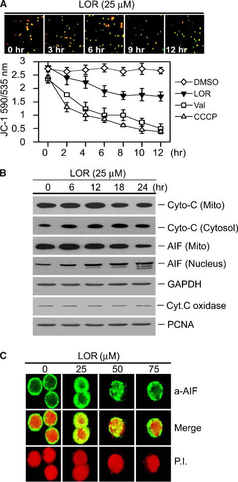
the Figure 5A and B, we found that LOR (25 mM)
the activation of caspase 3, we examined the changes
increased outer (cytochrome C and AIF release) and
of the protein levels of caspases 8 and 9 in the LOR-
inner (loss of DCm) mitochondria membrane per-
treated COLO 205 cells. Treatment of COLO 205 cells
meability. The release of cytochrome C and AIF were
with LOR (25–50 mM) resulted in caspase 9 activation
then observed as early as 6 h which kinetically
evidenced by degradation of the procaspase 9 but
paralleled a decreased of mitochondria membrane
not by caspase 8 because substrate of the caspase 8
potential (Figure 5A and B). These observations
(truncated form of Bid, t-Bid) was not degraded
suggest that LOR-induced apoptosis may be initiated
(Figure 6A). These observations suggest that LOR-
with early alterations in mitochondrial membrane
induced apoptosis is dependent on caspase 9 activa-
tion. Activation of caspase 9 in the Apaf-1 apopto-
As shown in the Figure 5B, the AIF was released
some is predominantly triggered by the release of
from mitochondria and translocated into nucleus as
cytochrome C from mitochondria into the cyto-
early as 6 h after LOR treatment in the COLO 205
plasm [56]. Accordingly, the cytosolic and mito-
cells. To our knowledge, AIF was demonstrated to be
chondrial fractions, prepared from cells treated with
involved in initiate the nuclear apoptotic events
LOR (25 mM) time-dependently were subjected to
including nuclear membrane degradation, periph-
Western blot analysis to assess the release of cyto-
eral chromatin condensation, large-scale fragmenta-
chrome C and demonstrated that the LOR induced a
tion of DNA, and, ultimately, cytotoxicity [55]. We
significant increase in cytosolic levels of cytochrome
then confirmed the LOR-induced nuclear transloca-
C as early as 6 h after LOR treatment (Figure 5B). To
tion of the AIF by immunofluorescence staining
further confirm the Western blot results, we perfor-
observed by confocal microscopic analysis. In con-
med caspase activity assays. As shown in Figure 6B,
sistent to the result shown in Figure 5B, our data
treatment of COLO 205 cells with 25 mM LOR indu-
demonstrated that AIF translocation to nucleus was
ced caspase 3 activity about 7.8-folds and 11.2-folds
found in COLO 205 cells at 6 h after LOR (25–50 mM)
at 9 and 12 h compared to control, respectively,
treatment. As shown in the Figure 5C, the AIF
while induced caspase 9 activity about 5.1-folds
translocation to nucleus was not observed in COLO
and 7.8-folds at 9 and 12 h compared to control,
205 cells after treated with higher dose of LOR
respectively. We therefore hypothesized that LOR-
(75 mM). Similar result was observed in the Figure 1B
mediated apoptosis was through the mitochondria
indicated that LOR-induced DNA fragmentation was
signaling pathways in which the caspase 9 protein
not observed in the COLO 205 cells when exposed to
may be activated.
higher dose (75 mM) LOR. Such observations indicat-
We next examined the therapeutic efficacy of LOR
ing that higher dose LOR may trigger some mediators
(25 mg/Kg) in vivo by treating athymic nude mice
which induced G2/M cell growth arrest effects and
bearing COLO 205 tumor xenografts. After establish-
may contribute to inhibit of cellular apoptosis stress
ment of palpable tumors (mean tumor volume,
200 mm3), animal received I.P. injections of LOR
To investigate the molecular events of LOR-
three times per wk, as well as DMSO plus peanut oil as
induced apoptosis, we then investigated the apo-
a vehicle control. After 6 wks, gross morphology of
Figure 5. Evaluation on the role of the mitochondria signals
AIF released form mitochondria into the cytoplasmic and nuclear
involved in LOR induced apoptosis. (A) The top insets, mitochondria
were then determined by Western blot analysis. The expression levels
membrane depolarization in LOR-treated COLO 205 cells was
of the cytochrome C oxidase (subunit IV), GAPDH, and PCNA
measured by JC-1 staining. In the lower panel, COLO 205 cells were
proteins were employed as a loading control to rule out cross
treated with LOR (25 mM), Val (200 mM), or with CCCP (200 mM) for
contamination of the mitochondria, cytoplasmic, and nuclear
the indicated time points. After drug treatment, the COLO 205 cells
fractions, respectively. (C) Confocal microscopic analysis for
were stained with JC-1 (1 mg/ml) as described in Materials and
nuclear/cytoplasmic distribution of AIF in control (DMSO-treated)
Methods. Results were expressed as a change in the ratio between
and LOR-treated cells. Cells were treated with DMSO (control) or LOR
red JC-1 fluorescence (Em 590 nm) and green JC-1 fluorescence (Em
(25–75 mM) for 4 h and then stained with anti-AIF antibody (green)
535 nm) over time. Each point represents the mean SE from three
or P.I. (red). [Color figure can be viewed in the online issue, which is
independent experiments. (B) COLO 205 cells were treated with LOR
available at www.interscience.wiley.com.]
(25 mM) time dependently. The protein level of the cytochrome C and
significance of application for cancer chemothera-peutic purposes.
LOR, which is a promising world widely used oral-
anti-histamine agent, has been used in the treatmentof allergic disease. In this study, we have shown thatLOR effectively inhibits proliferation of humancolon cancer (COLO 205) cells by causing G2/Mcell-cycle arrest and caspase 9-mediated apoptosis. Invivo study revealed that the growth of COLO 205xenografts in nude mice was retarded significantlyupon I.P. administration of LOR with a clinicaltherapeutic relevance dose (25 mg/kg). These resultsprompted us to examine further the mechanisms bywhich LOR inhibits proliferation of cancer cells.
Recent studies demonstrated that cells response to
DNA damage agents initiated two distinct check-point kinases namely Chk1 and Chk2 [18,57].
Recently, several Chk1 and Chk2 checkpoint abro-gating agent (abrogator) have been developed asadjuvants and intended to improve the therapeuticindex of cancer chemotherapy [23,58]. The originalstudies were performed by using caffeine, a non-specific G2 abrogator which disrupt the G2 check-point to sensitize G1-defective cancer cells intoapoptosis [59,60]. In our study, the Chk1 specificinhibitor (SB-218078) were used as a G2 phaseabrogator and demonstrated that SB-218078 abro-gate the LOR-induced G2/M arrest and sensitized thecells from G2/M arrest into apoptosis (Figure 4D).
Additional studies also demonstrated that knock-down of Chk1, Wee1, and Myt1 by RNA interferenceabrogates either adriamycin- or paclitaxel-mediatedG2 checkpoint and induces apoptosis in humancancer cells [17,58,61]. Therefore, Chk1 downregu-lation can not only potentiate DNA-damagingagents, but also enhance the toxicity of anti-micro-tubule agents (such as paclitaxel) [12,17,58], which
Figure 6. Dose-dependent response of caspase activity involved in
significantly broadens its therapeutic applications.
LOR-mediated apoptosis in COLO 205 cells. (A) COLO 205 cells were
In our study, the LOR-induced G2/M phase arrest
treated with LOR (10–75 mM) for 24 h and the expression levelsof caspase-associated proteins were determined by Western blot
was completely attenuated by SB218078 and DBH
analysis. The expression level of the GAPDH protein was selected and
but not by caffeine (Figure 4B). Caffeine has been
as a loading control. (B) COLO 205 cells were treated with 25 mM
shown to inhibit the activities of both ATM and ATR
LOR for the indicated time points. The LOR-induced caspase activitiesin COLO 205 cells were measured as described in Materials and
upstream checkpoint kinases and to override the
Methods. Data was represented as mean SE from three indepen-
ATM- and ATR-dependent DNA damage checkpoints
[49]. Recently, a cell-free experimental system fromXenopus eggs extracts was performed and described a
the tumor volume in mice treated with LOR (Figure
caffeine-insensitive pathway which plays some
7A, mice No. 5–8) was significant smaller in
important role in the checkpoint response [62,63].
comparison with DMSO-treated controls (Figure
Similar reports in human colon cancer cells also
7A, mice No. 1–4). A reduction in tumor volume
demonstrated that ATR-mediated DNA-damage
between mice given LOR versus those given vehicle
checkpoint was not completely rescued by caffeine.
(DMSO) was detected (Figure 7B–E). In mice receiv-
For example, increased phosphorylation of gH2AX
ing these treatment regimens, no gross signs of
was regulated by ATR in response to DNA breaks
toxicity were observed (body weight, visible inspec-
observed in human colon cancer cells [64]. The
tion of general appearance, and microscopic exam-
phosphorylated gH2AX was also induced in human
ination of individual organs). Our results provide
colon cancer (HCT 116) cells by DNA alkylating
further evidences that such observations may have
agent, hedamycin. In this study, caffeine did not
ANTITUMOR EFFECTS OF LOR IN HUMAN COLON CANCER CELLS GROWTH
Figure 7. The growth of COLO 205 tumor xenografts in nude
(shown as No. 5–8) for 6 wk. (B) Average tumor volume of DMSO-
mice was retarded by LOR treatment. Athymic nude mice injected
treated (circle) versus LOR-treated (square) nude mice, (C) body
with COLO 205 cells into subcutaneous tissue of inter-scapular area.
weight, (D) tumor weight, and (E) tumor/body weight ratio were
Once tumor volume reached approximately 200 mm3, the animal
measured at the end of the experiment. Four samples were analyzed
received treatment of 25 mg/kg LOR, or DMSO intraperitoneally
in each group and values represent the mean SE. Comparisons
three times per wk for 6 wk. (A) Gross appearance of subcutaneous
were subjected to Student's t test. *P < 0.05 versus control.
tumors after treatment with DMSO (shown as No. 1–4) or LOR
substantially reduce induction of H2AX phosphor-
not completely reverse the LOR-induced G2/M arrest
ylation by hedamycin [65]. The H2AX phosphoryla-
(Figure 4C). These results suggest that a caffeine-
tion was also induced by silibinin, a naturally
sensitive pathway responsible for maintenance of
occurring flavonoid, which against ultraviolet B
the LOR-induced G2/M-arrested cells is dependent
(UVB)-induced skin tumorigenesis. However, the
on both ATR and Chk1 checkpoint kinases. In such a
anti-tumor effects of silibinin is possibly not through
situation, the G2/M arrest induced by LOR can be
ATM/ATR activation, as caffeine pretreatment had
prevented by caffeine pretreatment. However, our
no effect on silibinin plus UVB induced p53-Ser15
data indicated that at least part of LOR-induced G2/M
arrested cells utilize a caffeine-insensitive pathway,
As described above, caffeine is a well-known
that is, that checkpoint signaling by ATM or ATR is
inhibitor of both ATM and ATR pathways, it could
most likely not involved. This result cannot exclude
a possibility indicated that a distinct, caffeine-
the residue of Ser-155 [46], which results in seques-
insensitive ATM/ATR signaling pathway might also
tering BAD from mitochondria into cytosol by
be necessary to attenuate the effects of LOR-
interacting with 14-3-3 [75]. The LOR-mediated
mediated G2/M arrest and also plays a role in the
Chk1 kinase activity was inhibited by SB-218078
which prevent the formation of p-BAD/14-3-3 com-
In our and others previous studies [12,26,41],
plex and promoted the G2/M phase arrested cells into
the cyclin B1 protein was induced in the microtubule
apoptosis in the COLO 205 cells (Figure 4A).
damaging agents-mediated G2/M arrested cells due
In summery, the results of this study provide
to block of the mitotic process. However, decreased
evidence that LOR-induced G2/M arrest in COLO
of cyclin B1 protein level was also observed in the
205 cells involves regulation of Cdc2 activity
G2/M arrested cells through inhibition of the mRNA
through three distinct mechanisms, which include
synthesis and accelerate degradation of the cyclin B1
LOR-induced activation of Chk1 kinase and LOR-
protein [5,67–69]. The cyclin B1 suppression prob-
induced downregulation of Cdc25C protein levels.
ably prevents the optimal formation of the mitosis-
Each of these mechanisms results in a decrease in
or maturation-promoting factor (MPF) which con-
Cdc2 kinase activity. Of particular interest is the
sists of a complex between the cyclin B1 and the
finding that the LOR-induced activation of Chk1 is
Cdc2 kinase protein, and is essential for cells to cross
implicated in each of these mechanisms of Cdc2
the threshold from G2 into mitosis. In this study, our
regulation. Thus, studies in this report indicate that
results revealed that cyclin B1 inhibition and its
activation of Chk1 plays a central role, either directly
inactivation of the cyclinB1/Cdc2 kinase activity
or indirectly, in the induction of G2/M cell-cycle
may be play an essential role in LOR-induced G2/M
arrest and apoptosis by LOR through alterations in
arrest. Such hypothesis has also been proposed in
Cdc25C and BAD protein localization and activity.
previous studies revealed that cyclin B1 expression
However, our study still needed to address a funda-
in human cancer cells was inhibited by different
mental question, which remains unanswered, is how
stimulations including ionizing radiation [70], DNA
LOR treatment causes DNA damage to activate ATR/
damaging agents [67], or cancer chemopreventive
Chk1. Additional studies needed to identify the
agents [5,71,72]. In addition, recent studies have
specific mechanisms of LOR-mediated activation of
shown that ectopic expression of BRCA1 in human
Chk1, as well as the precise role of Chk1 on the
cells can trigger cellular responses including G2/M
regulation of G2/M arrest and apoptosis.
cell cycle arrest and apoptosis [68,69]. The BRCA1regulates the G2/M checkpoint was also demon-
strated by activating Chk1 kinase and inhibition ofcyclin B1 upon DNA damage [69]. Similarly, our
This study was supported by the National Science
findings of decreased cyclin B1 expression concomi-
Council grant NSC 92-2314-B-038-029 to Dr. Ho, and
tant with the induction of Chk1 in the LOR-induced
NSC 92-2320-B-038-018 to Dr. Lee.
G2/M arrested cells were in agreement with the datafrom Yarden et al. (2002) [69].
As described above, the Cdc25C phosphatase is a
dual specific phosphatase that triggers the activation
1. Hunter T, Pines J. Cyclins and cancer II: Cyclin D and CDK
of Cyclin B1/Cdc2 by dephosphorylating Cdc2 at
inhibitors come of age. Cell 1994;79:573–582.
the residues Thr-14 and Tyr-15 [73]. The activity of
2. Huang TS, Shu CH, Chao Y, Chen SN, Chen LL. Activation of
Cdc25C is downregulated by Chk1 kinase through
MAD 2 checkprotein and persistence of cyclin B1/CDC 2activity associate with paclitaxel-induced apoptosis in human
phosphorylation of residue at Ser-216, which affects
nasopharyngeal carcinoma cells. Apoptosis 2000;5:235–
both the enzyme activity and the subcellular locali-
zation of the Cdc25C [74]. Our results indicated that
3. Kharbanda S, Yuan ZM, Taneja N, Weichselbaum R,
increased phosphorylation of Cdc25C (Ser-216) in
Kufe D. p56/p53lyn tyrosine kinase activation in mammalian
response to LOR treatment in the COLO 205 cells
cells treated with mitomycin C. Oncogene 1994;9:3005–3011.
was associated with Chk1 activation (Figure 4A). Our
4. Kawabe T, Suganuma M, Ando T, Kimura M, Hori H,
results shown in the Figure 3C–E confirmed the
Okamoto T. Cdc25C interacts with PCNA at G2/M transition.
previous reports suggested that the Cdc25C is phos-
phorylated by Chk1 kinase at the Ser-216 residue,
5. Singh SV, Herman-Antosiewicz A, Singh AV, et al. Sulfor-
aphane-induced G2/M phase cell cycle arrest involves
which generates consensus binding sites for 14-3-3
checkpoint kinase 2-mediated phosphorylation of cell
proteins and then inactivate the Cdc25C by seques-
division cycle 25C. J Biol Chem 2004;279:25813–25822.
trate this protein into the cytoplasm [5,69]. Thus,
6. Advani SJ, Brandimarti R, Weichselbaum RR, Roizman B. The
LOR-induced activation of Chk1 is important for the
disappearance of cyclins A and B and the increase in activity
subsequent inactivation of Cdc25C and Cdc2, which
of the G(2)/M-phase cellular kinase cdc2 in herpes simplexvirus 1-infected cells require expression of the alpha22/
eventually caused the G2/M cell-cycle arrest. The
U(S)1.5 and U(L)13 viral genes. J Virol 2000;74:8–15.
Chk1 has also been demonstrated to inactivate the
7. Dunphy WG. The decision to enter mitosis. Trends Cell Biol
proapoptotic protein BAD by phosphorylation at
ANTITUMOR EFFECTS OF LOR IN HUMAN COLON CANCER CELLS GROWTH
8. Wilker EW, Grant RA, Artim SC, Yaffe MB. A structural basis
mice through induction of apoptosis and G0/G1 cell cycle
for 14-3-3 sigma functional specificity. J Biol Chem 2005;
arrest. Toxicol Appl Pharmacol 2002;180:22–35.
28. Palermo C, Walworth NC. Assaying cell cycle checkpoints:
9. Peng CY, Graves PR, Ogg S, et al. C-TAK1 protein kinase
Activity of the protein kinase Chk1. Methods Mol Biol 2005;
phosphorylates human Cdc25C on serine 216 and promotes
14-3-3 protein binding. Cell Growth Differ 1998;9:197–
29. Zhao B, Bower MJ, McDevitt PJ, et al. Structural basis for
Chk1 inhibition by UCN-01. J Biol Chem 2002;277:46609–
10. Jiang K, Pereira E, Maxfield M, Russell B, Goudelock DM,
Sanchez Y. Regulation of Chk1 includes chromatin associa-
30. McGowan CH, Russell P. Human Wee1 kinase inhibits cell
tion and 14-3-3 binding following phosphorylation on Ser-
division by phosphorylating p34cdc2 exclusively on Tyr15.
345. J Biol Chem 2003;278:25207–25217.
EMBO J 1993;12:75–85.
11. Lee J, Kumagai A, Dunphy WG. Positive regulation of Wee1
31. McGowan CH, Russell P. Cell cycle regulation of human
by Chk1 and 14-3-3 proteins. Mol Biol Cell 2001;12:551–
WEE1. EMBO J 1995;14:2166–2175.
32. Yan Y, Mumby MC. Distinct roles for PP1 and PP2A in
12. Xiao Z, Xue J, Sowin TJ, Rosenberg SH, Zhang H. A novel
phosphorylation of the retinoblastoma protein. PP2a reg-
mechanism of checkpoint abrogation conferred by Chk1
ulates the activities of G cyclin-dependent kinases. J Biol
13. Shao RG, Cao CX, Pommier Y. Abrogation of Chk1-
33. Ho YS, Wang YJ, Lin JK. Induction of p53 and p21/WAF1/
mediated S/G2 checkpoint by UCN-01 enhances ara-C-
CIP1 expression by nitric oxide and their association with
induced cytotoxicity in human colon cancer cells. Acta
apoptosis in human cancer cells. Mol Carcinog 1996;16:
Pharmacol Sin 2004;25:756–762.
14. Suganuma M, Kawabe T, Hori H, Funabiki T, Okamoto T.
34. Ho YS, Ma HY, Chang HY, et al. Lipid peroxidation and cell
Sensitization of cancer cells to DNA damage-induced cell
death mechanisms in rats and human cells induced by chloral
death by specific cell cycle G2 checkpoint abrogation. Cancer
hydrate. Food Chem Toxicol 2003;41:621–629.
35. Lin SY, Chang YT, Liu JD, et al. Molecular mechanisms of
15. Xiao Z, Xue J, Semizarov D, Sowin TJ, Rosenberg SH, Zhang
apoptosis induced by magnolol in colon and liver cancer cells.
H. Novel indication for cancer therapy: Chk1 inhibition
Mol Carcinog 2001;32:73–83.
sensitizes tumor cells to antimitotics. Int J Cancer 2005;115:
36. Maianski NA, Geissler J, Srinivasula SM, Alnemri ES, Roos D,
Kuijpers TW. Functional characterization of mitochondria in
16. Jin ZH, Kurosu T, Yamaguchi M, Arai A, Miura O.
neutrophils: A role restricted to apoptosis. Cell Death Differ
Hematopoietic cytokines enhance Chk1-dependent G2/M
checkpoint activation by etoposide through the Akt/GSK3
37. Liu JD, Wang YJ, Chen CH, et al. Molecular mechanisms of
pathway to inhibit apoptosis. Oncogene 2005;24:1973–
G0/G1 cell-cycle arrest and apoptosis induced by terfenadine
in human cancer cells. Mol Carcinog 2003;37:39–50.
17. Ren Q, Liu R, Dicker A, Wang Y. CHK1 affects cell sensitivity
38. Wang YJ, Yu CF, Chen LC, et al. Ketoconazole potentiates
to microtubule-targeted drugs. J Cell Physiol 2005;203:
terfenadine-induced apoptosis in human Hep G2 cells
through inhibition of cytochrome p450 3A4 activity. J Cell
18. Huang X, Tran T, Zhang L, Hatcher R, Zhang P. DNA damage-
induced mitotic catastrophe is mediated by the Chk1-
39. Grant JA, Danielson L, Rihoux JP, DeVos C. A double-blind,
dependent mitotic exit DNA damage checkpoint. Proc Natl
single-dose, crossover comparison of cetirizine, ebastine,
Acad Sci USA 2005;102:1065–1070.
epinastine, fexofenadine, terfenadine, and loratadine versus
19. Sancar A, Lindsey-Boltz LA, Unsal-Kacmaz K, Linn S. Molec-
placebo: Suppression of histamine-induced wheal and flare
ular mechanisms of mammalian DNA repair and the DNA
response for 24 h in healthy male subjects. Allergy 1999;54:
damage checkpoints. Annu Rev Biochem 2004;73: 39–85.
20. Liu Q, Guntuku S, Cui XS, et al. Chk1 is an essential kinase
40. Nyberg KA, Michelson RJ, Putnam CW, Weinert TA. Toward
that is regulated by Atr and required for the G(2)/M DNA
maintaining the genome: DNA damage and replication
damage checkpoint. Genes Dev 2000;14:1448–1459.
checkpoints. Annu Rev Genet 2002;36:617–656.
21. Jackson JR, Gilmartin A, Imburgia C, Winkler JD, Marshall LA,
41. Tseng CJ, Wang YJ, Liang YC, et al. Microtubule damaging
Roshak A. An indolocarbazole inhibitor of human checkpoint
agents induce apoptosis in HL 60 cells and G2/M cell cycle
kinase (Chk1) abrogates cell cycle arrest caused by DNA
arrest in HT 29 cells. Toxicology 2002;175:123–142.
damage. Cancer Res 2000;60:566–572.
42. Guo Z, Kumagai A, Wang SX, Dunphy WG. Requirement for
22. Hutchins JR, Dikovskaya D, Clarke PR. Regulation of Cdc2/
Atr in phosphorylation of Chk1 and cell cycle regulation in
cyclin B activation in Xenopus egg extracts via inhibitory
response to DNA replication blocks and UV-damaged DNA in
phosphorylation of Cdc25C phosphatase by Ca(2þ)/calmo-
Xenopus egg extracts. Genes Dev 2000;14:2745–2756.
dulin-dependent protein [corrected] kinase II. Mol Biol Cell
43. Sanchez Y, Wong C, Thoma RS, et al. Conservation of the
Chk1 checkpoint pathway in mammals: Linkage of DNA
23. Kawabe T. G2 checkpoint abrogators as anticancer drugs.
damage to Cdk regulation through Cdc25. Science 1997;
Mol Cancer Ther 2004;3:513–519.
24. Siddiqui KM, Chopra DP. Primary and long term epithelial cell
44. Furnari B, Rhind N, Russell P. Cdc25 mitotic inducer targeted
cultures from human fetal normal colonic mucosa. In Vitro
by chk1 DNA damage checkpoint kinase. Science 1997;277:
25. Lee WS, Chen RJ, Wang YJ, et al. In vitro and in vivo studies of
45. Matsuoka S, Huang M, Elledge SJ. Linkage of ATM to cell
the anticancer action of terbinafine in human cancer cell
cycle regulation by the Chk2 protein kinase. Science 1998;
lines: G0/G1 p53-associated cell cycle arrest. Int J Cancer
46. Han EK, Butler C, Zhang H, et al. Chkl binds and phospho-
26. Ho YS, Duh JS, Jeng JH, et al. Griseofulvin potentiates
rylates BAD protein. Anticancer Res 2004;24:3907–3910.
antitumorigenesis effects of nocodazole through induction
47. Masters SC, Yang H, Datta SR, Greenberg ME, Fu H. 14-3-3
of apoptosis and G2/M cell cycle arrest in human colorectal
inhibits Bad-induced cell death through interaction with
cancer cells. Int J Cancer 2001;91:393–401.
serine-136. Mol Pharmacol 2001;60:1325–1331.
27. Wu CH, Jeng JH, Wang YJ, et al. Antitumor effects of
48. Curman D, Cinel B, Williams DE, et al. Inhibition of the G2
miconazole on human colon carcinoma xenografts in nude
DNA damage checkpoint and of protein kinases Chk1 and
Chk2 by the marine sponge alkaloid debromohymenialdi-
62. Kumagai A, Guo Z, Emami KH, Wang SX, Dunphy WG. The
sine. J Biol Chem 2001;276:17914–17919.
Xenopus Chk1 protein kinase mediates a caffeine-sensitive
49. Sarkaria JN, Busby EC, Tibbetts RS, et al. Inhibition of ATM
pathway of checkpoint control in cell-free extracts. J Cell Biol
and ATR kinase activities by the radiosensitizing agent,
caffeine. Cancer Res 1999;59:4375–4382.
63. Luciani MG, Oehlmann M, Blow JJ. Characterization of a
50. Zhou BB, Chaturvedi P, Spring K, et al. Caffeine abolishes the
novel ATR-dependent, Chk1-independent, intra-S-phase
mammalian G(2)/M DNA damage checkpoint by inhibiting
checkpoint that suppresses initiation of replication in
ataxia-telangiectasia-mutated kinase activity. J Biol Chem
Xenopus. J Cell Sci 2004;117:6019–6030.
64. Bartkova J, Horejsi Z, Koed K, et al. DNA damage response as
51. Blasina A, Price BD, Turenne GA, McGowan CH. Caffeine
a candidate anti-cancer barrier in early human tumorigen-
inhibits the checkpoint kinase ATM. Curr Biol 1999;9:1135–
esis. Nature 2005;434:864–870.
65. Tu LC, Matsui SI, Beerman TA. Hedamycin, a DNA alkylator,
52. Wang YJ, Jeng JH, Chen RJ, et al. Ketoconazole potentiates
induces (gamma)H2AX and chromosome aberrations: Invol-
the antitumor effects of nocodazole: In vivo therapy for
vement of phosphatidylinositol 3-kinase-related kinases and
human tumor xenografts in nude mice. Mol Carcinog 2002;
DNA replication fork movement. Mol Cancer Ther 2005;4:
53. Godlewski MM, Gajkowska B, Lamparska-Przybysz M, Motyl
66. Dhanalakshmi S, Agarwal C, Singh RP, Agarwal R. Silibinin
T. Colocalization of BAX with BID and VDAC-1 in nimesulide-
up-regulates DNA-protein kinase-dependent p53 activation
induced apoptosis of human colon adenocarcinoma COLO
to enhance UVB-induced apoptosis in mouse epithelial JB6
205 cells. Anticancer Drugs 2002;13:1017–1029.
cells. J Biol Chem 2005;280:20375–20383.
54. Godlewski MM, Motyl MA, Gajkowska B, Wareski P,
67. Manni I, Mazzaro G, Gurtner A, et al. NF-Y mediates the
Koronkiewicz M, Motyl T. Subcellular redistribution of BAX
transcriptional inhibition of the cyclin B1, cyclin B2, and
during apoptosis induced by anticancer drugs. Anticancer
cdc25C promoters upon induced G2 arrest. J Biol Chem
55. Hong SJ, Dawson TM, Dawson VL. Nuclear and mitochon-
68. Yan Y, Spieker RS, Kim M, Stoeger SM, Cowan KH. BRCA1-
drial conversations in cell death: PARP-1 and AIF signaling.
mediated G2/M cell cycle arrest requires ERK1/2 kinase
Trends Pharmacol Sci 2004;25:259–264.
activation. Oncogene 2005.
56. Qin H, Srinivasula SM, Wu G, Fernandes-Alnemri T, Alnemri
69. Yarden RI, Pardo-Reoyo S, Sgagias M, Cowan KH, Brody LC.
ES, Shi Y. Structural basis of procaspase-9 recruitment by
BRCA1 regulates the G2/M checkpoint by activating Chk1
the apoptotic protease-activating factor 1. Nature 1999;399:
kinase upon DNA damage. Nat Genet 2002;30:285–289.
70. Theron T, Bohm L. Cyclin B1 expression in response to
57. Ho CC, Siu WY, Chow JP, et al. The relative contribution of
abrogation of the radiation-induced G2/M block in HeLa
CHK1 and CHK2 to Adriamycin-induced checkpoint. Exp Cell
cells. Cell Prolif 1998;31:49–57.
71. Brown JW, Cappell S, Perez-Stable C, Fishman LM. Extracts
58. Xiao Z, Xue J, Semizarov D, Sowin TJ, Rosenberg SH, Zhang
from two marine sponges lower cyclin B1 levels, cause a G2/
H. Novel indication for cancer therapy: Chk1 inhibition
M cell cycle block and trigger apoptosis in SW-13 human
sensitizes tumor cells to antimitotics. Int J Cancer 2005.
adrenal carcinoma cells. Toxicon 2004;43:841–846.
59. Powell SN, DeFrank JS, Connell P, et al. Differential sensitivity
72. Scaife RM. G2 cell cycle arrest, down-regulation of cyclin B,
of p53() and p53(þ) cells to caffeine-induced radio-
and induction of mitotic catastrophe by the flavoprotein
sensitization and override of G2 delay. Cancer Res 1995;
inhibitor diphenyleneiodonium. Mol Cancer Ther 2004;3:
60. Russell KJ, Wiens LW, Demers GW, Galloway DA, Plon SE,
73. Bulavin DV, Higashimoto Y, Demidenko ZN, et al. Dual
Groudine M. Abrogation of the G2 checkpoint results in
phosphorylation controls Cdc25 phosphatases and mitotic
differential radiosensitization of G1 checkpoint-deficient
entry. Nat Cell Biol 2003;5:545–551.
and G1 checkpoint-competent cells. Cancer Res 1995;55:
74. Smits VA, Medema RH. Checking out the G(2)/M transition.
Biochim Biophys Acta 2001;1519:1–12.
61. Wang Y, Decker SJ, Sebolt-Leopold J. Knockdown of Chk1,
75. Hsu SY, Kaipia A, Zhu L, Hsueh AJ. Interference of BAD (Bcl-
Wee1 and Myt1 by RNA interference abrogates G2
xL/Bcl-2-associated death promoter)-induced apoptosis in
checkpoint and induces apoptosis. Cancer Biol Ther 2004;
mammalian cells by 14-3-3 isoforms and P11. Mol Endocrinol
Source: http://mts.tmu.edu.tw/BreastCancer/files/writing_journal/4/23_465fc951.pdf
herausgegeben von der Stelle für Presse und Öffentlichkeitsarbeit des Bundespatentgerichts Hausanschrift: Cincinnatistraße 64 Internet: www.bpatg.de E-Mail: [email protected] Schwerpunkt auch dieses Jahresberichts ist die Dokumentation der Rechtsprechung des Bundespatentgerichts. In den Bereichen Patentrecht, Gebrauchsmusterrecht, Geschmacksmusterrecht und Markenrecht werden insbesondere die Entscheidungen
This Accepted Manuscript has not been copyedited and formatted. The final version may differ from this version. Research Article: New Research Sensory and Motor Systems Effects of fluoxetine and visual experience on glutamatergic andGABAergic synaptic proteins in adult rat visual cortex Effects of fluoxetine in adult rat V1 Simon Beshara , Brett R Beston