Perturbation of estrogen receptor α localization with synthetic nona-arginine lxxll- peptide coactivator binding inhibitors

Chemistry & Biology Perturbation of Estrogen Receptor a Localizationwith Synthetic Nona-Arginine LXXLL-Peptide Coactivator Binding Inhibitors Mae¨lle Carraz,Wilbert Zwart,Trang Rob Michalides,and Luc 1Chemical Genomics Centre of the Max Planck Society, 44227 Dortmund, Germany2Laboratory of Chemical Biology, Department of Biomedical Engineering, Eindhoven University of Technology, 5600MB Eindhoven,The Netherlands3Division of Cell Biology, The Netherlands Cancer Institute, 1066CX Amsterdam, The Netherlands*Correspondence: DOI 10.1016/j.chembiol.2009.06.009 coactivators includes the p160 protein family, consisting of threemembers: SRC-1, Tif-2/GRIP1, and SRC-3/AIB1. The impor- The interaction of estrogen receptor a (ERa) with the tance of these coactivators in ERa signaling has been high- consensus LXXLL motifs of transcriptional coactiva- lighted by knockout studies and by the discovery of p160 gene tors provides an entry for functional ERa inhibi- amplification in ERa-positive breast cancer tion. Here, synthetic cell-permeable LXXLL peptide These p160 coactivators probes are brought forward that allow evaluation of have the ability to interact with ERa and other nuclear receptors the interaction of specific recognition motifs with in a hormone-dependent manner via small hydrophobic andamphiphatic a-helical peptide sequences with the common ERa in the context of the cell. The probes feature a signature motif, LXXLL (L = leucine, and X = any amino acid).
nona-arginine tag that facilitates cellular entry and Crystallographic studies showed that this motif, also called induces probe localization in nucleoli. The nucleoli nuclear receptor box (NR box), binds to a hydrophobic cleft on localization provides an explicit tool for evaluating the ERa ligand binding domain (LBD) surface, under control of the LXXLL motif interaction with ERa. The probes activating ligand (p160 coactivators contain compete with coactivators, bind ERa, and recruit it three NR box copies within their central nuclear receptor interac- into the nucleoli. The physical inhibition of the ERa- tion domain. Although the three leucines in the LXXLL motif are coactivator interaction by the probes is shown to conserved, the amino acid residues flanking this sequence are be correlated with the inhibition of ERa-mediated not and specify NR box recognition for a particular nuclear gene transcription. This chemical biology approach receptor in cooperation with specificities of the ligand. These allows evaluating the ER characteristic features have raised the idea that the ERa-coacti- vator interaction is a specific amendable interface for LXXLL and inhibitor binding directly in cells.
motif–like inhibitors, preventing transactivation by ERa The formation of the coactivator binding surface on ERa requires a hormone-dependent conformational change. Inhibitors of coactivator Estrogen receptor a (ERa) is a member of the nuclear receptor binding (ICBs) may only bind to superfamily and is an estrogen-inducible transcription factor. In hormone-occupied ERa, thus featuring a different mechanism the normal physiology of the female reproductive system, ERa than the classic anti-estrogen drugs. These ICBs might thus be regulates the growth and function of such tissues as breast, useful in anti-estrogen therapy for patients with tamoxifen-resis- uterus, and ovaries. In addition, ERa has been identified as play- tant breast cancers or could provide a possibility to target ing a role in many pathological processes, such as breast cancer orphan nuclear receptors for which antagonists are difficult to (). Because 70% of breast tumors depend on the stimulatory effect of estrogens, anti-estrogen therapies Several types of compounds blocking ERa-cofactor interac- have been developed that significantly reduce breast cancer tions have already been developed. The first proof that the recurrence ). However, the long-term effects concept of inhibiting ERa-cofactor interactions results in the inhi- of tamoxifen, one of the most successful anti-estrogen drugs, bition of ERa-mediated transcription has been obtained with in metastatic disease are limited to an average of 15 months LXXLL peptides overexpressed in cells from transfected DNA (), after which tamoxifen resistance develops in more than 80% of treated women In the induction of gene transcription, ERa acts in complex association has been enforced by the identification and the use of synthetic with other cellular factors, including coactivators, which are ICBs, either peptide based or small molecule required for the transcriptional activation process to occur (The most widely studied group of ERa 702 Chemistry & Biology 16, 702–711, July 31, 2009 ª2009 Elsevier Ltd All rights reserved Chemistry & Biology Cellular Modulation of ERa with Synthetic Peptides Most of these compounds inhibit Table 1. Sequences of the Selected LXXLL-Motif Peptides ERa-coactivator binding in biochemical assays, and a selected set of compounds was also able to inhibit ERa gene transactiva- tion in cellular assays ). The translation from biochemical studies on the ERa LBD to cellular activity on the full-length protein is, however, not trivial. For example, it has been reported that the conformation of the coactivator binding groove in the full-length receptor tends to be different from the isolated LBD ( addition, direct binding of inhibitors to ERa in cells and their capacity to displace coactivators are two processes that aredifficult to observe directly. The process of inhibiting the ERa-co-activator interaction thus requires more molecular insight. There- complete peptide. In addition to the LXXLL molecular probes, fore, methods need to be developed that directly visualize the two AXXAA-molecular probes, based on SRC-1 Box2 and Tif-2 interaction of inhibitors with ERa and provide information on Box2 and mutated for the recognition motif by alanines, were the resulting ERa inhibition. Here, we show that synthetic cell- also generated as negative control peptides.
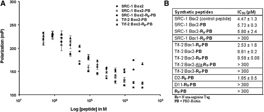
permeable LXXLL-peptide probes with specific localization and The effect of the biotin and nona-arginine tags, incorporated in visualization tags are ideal molecular probes to inhibit and eval- the peptides, on the binding to the ERa coactivator binding uate ERa-coactivator interactions in living cells and to address surface was evaluated in a biochemical fluorescent polarization the above-mentioned issues. By use of confocal laser scanning assay with the recombinant LBD of ERa The assay microscopy (CLSM), these probes can be used to monitor the measures the displacement of a fluorescein-labeled reference association of specific peptide sequences with the full-length peptide (SRC-1 box2) by the nonfluorescent LXXLL peptides ERa and to evaluate their effects on the assembly of ERa under study. The R9 tag alone did not bind to ERa LBD, as signaling protein complex within the organized microenviron- expected. The dose-dependent fluorescence depolarization ment of the living cell. The peptide probes feature a nona-argi- curves obtained with peptides SRC-1 Box2 and SRC-1 Box2- nine tag that ensures efficient cellular entry of the peptides and PEG-biotin were overlapping A), showing that the induces a specific localization of the peptides into the nucleoli.
PEG-biotin spacer does not influence ERa binding. For both This specific property of the peptides is used to evaluate the the SRC-1 Box2 and the SRC-1 Box2-R9 peptides and the Tif- affinity of different LXXLL-motif peptides for the ERa. The 2 Box2 and Tif-2 Box2-R9 peptides, the same binding affinities peptides bind and translocate ERa into the nucleoli, resulting were found B), showing that the nona-arginine tag in the displacement of ERa from the DNA and coactivators.
also does not disturb the capacity of the peptides to bind to ERa.
Overall, the peptides function as functional inhibitors as theyeffectively lower ERa-mediated gene transcription.
Cellular Toxicity and Intracellular Distributionof the Synthetic LXXLL Molecular Probes To study and evaluate any toxic effects of the synthetic peptideprobes, the viability of cells after an overnight incubation with Selection and Synthesis of LXXLL-Motif Molecular increasing concentrations of peptides was tested using the WST-1 assay (see available online) ( For the generation of molecular probes inhibiting the ERa-coac- ). Peptides conjugated to PEG-biotin but lacking the tivator interaction, we selected a set of natural LXXLL-motif nona-arginine tag were found to be toxic on the human cancer sequences reported to have a high affinity for ERa cell line U2OS at concentrations of 1 mM, whereas all of their and capable of blocking ERa transcriptional activity nona-arginine counterparts showed toxicity at around 0.1 mM.
when overexpressed in cells—boxes 1 and 2 of the SRC-1 coac- The nona-arginine tag alone was found to be less toxic to the tivator protein and boxes 1, 2, and 3 of the Tif-2 coactivator cells. Interestingly, the two peptides SRC-1 Box2-Ala-R9 and protein (). Phage display peptides D2 and D11, which Tif-2 Box2-Ala-R9 mutated for the recognition motif by alanines were previously reported to possess a high affinity for full-length were not lethal to cells at concentrations up to 0.6 mM. This ERa and to bind at the same coactivator binding surface ( difference in minimal toxic concentration may be explained ), were also included in the selection The by a different sequence-dependent cellular uptake capacity of plasma membrane of mammalian cells is generally impermeable these peptides, but may also be related to their intracellular phar- to the vast majority of peptides and proteins. We therefore macological effects. From the dose-dependent toxicity curves decided to take advantage of the cell-permeable properties of obtained, we determined the concentrations of peptides that the nona-arginine (R9) peptide sequence gave less than 20% of cell death, compared with nontreated ) and use it as a carrier for the selected cells (). These peptide concentrations (0.02–0.07 mM), LXXLL peptides. The arginine tags were attached to the peptides were also tested by fluorescent assisted cell sorting (FACS) for at their C terminus by means of peptide synthesis. To monitor the apoptosis induction using the specific fluorescently labeled localization of the LXXLL peptides within the cells via immuno- apoptotic marker Annexin-V (). Also in this labeling, they were provided with a biotin epitope connected assay (data not shown), the nona-arginine peptides induced via an ethylene glycol linker to the C-terminal part of the less than 20% apoptosis at these concentrations.
Chemistry & Biology 16, 702–711, July 31, 2009 ª2009 Elsevier Ltd All rights reserved 703


Chemistry & Biology Cellular Modulation of ERa with Synthetic Peptides Figure 1. Biochemical Fluorescence Polari-zation Studies of the ERa-LBD BindingCapacity of the Synthetic Peptides(A) Representative dose-dependent inhibitioncurves of selected peptides for the binding ofa fluorescein-labeled SRC-1 Box2 peptide to theERa-LBD.
(B) IC50 values of all tested peptides. Error barsrepresent mean ± standard error.
The cell-entry and intracellular distributions of the synthetic zinc-finger motifs present in its DNA binding domain. Inhibiting peptides were assessed by immunolabeling of cells treated peptides expressed from DNA constructs fused to a fluorescent with the peptides using a fluorescent Cy5-conjugated anti-biotin protein are evenly distributed throughout the nucleus, but also in antibody. CLSM imaging of these cells revealed that the PEG- the cytoplasm, and are colocalized with ERa upon hormone biotin spacer and the LXXLL peptides lacking a nona-arginine addition We investigated here whether tag did cross the plasma membrane to some extent but clus- peptide probes of a synthetic nature and featuring a specific tered into vesicular structures in the cytoplasm ).
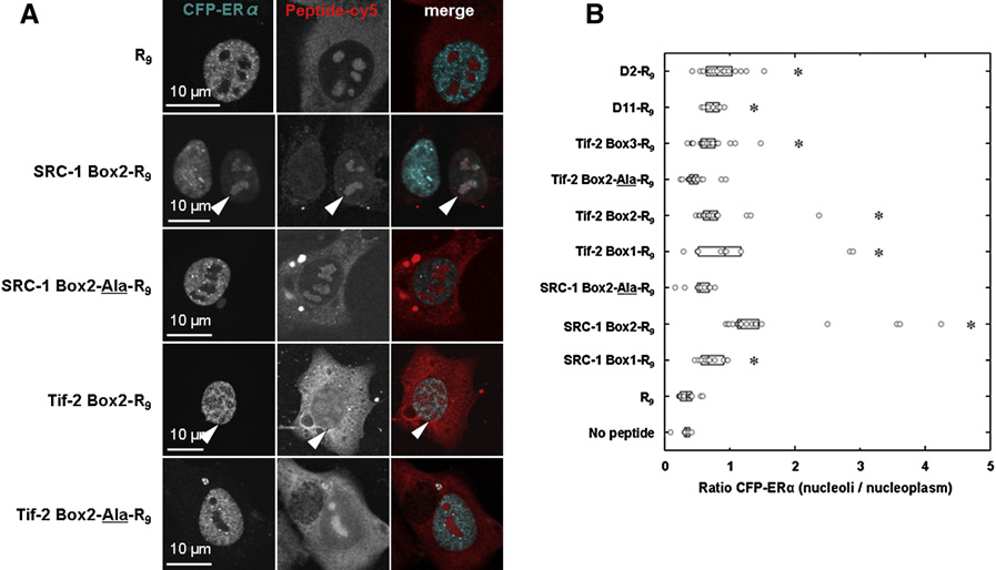
localization tag are able to interact with ERa as well, when However, the cellular distribution of the nona-arginine-tagged applied externally to cells, and whether the introduced localiza- molecular probes was completely different, localizing in both tion tag could be used to evaluate this interaction in a novel the cytoplasm and the nucleus. In the nucleus, these peptides manner. Therefore, U2OS cells transiently transfected with full- were mainly found located at the nucleoli, as anticipated length ERa fused to cyan fluorescent protein (CFP) were incu- () because the nucleolar accumulation of such positively bated with the LXXLL-nona-arginine molecular probes in the charged peptides has been described elsewhere presence of estradiol. The ERa-binding capacity of these Because this particular localization at the nucleoli peptides was assessed by colocalization with ERa by CLSM.
was not found for the non-R9-tagged peptides, we conclude When cells were not incubated with the molecular probes, ERa that this feature can be exclusively ascribed to the R9 tag.
localized as expected in the nucleoplasm, in punctuated foci Also, in accordance with the results of a recent study (data not shown). The same ERa distribution was observed in ), we observed that the concentration of serum presence of the nona-arginine tag alone, whereas this peptide used in the culture medium had a significant impact on the distri- itself was clearly observed into the nucleoli (indicating bution of these molecular probes inside cells (data not shown). In a lack of interaction between the receptor and this peptide.
addition, we noticed that supplementing the medium with 10% However, incubating the cells with the peptides SRC-1 Box2- of non-heat-inactivated serum could abrogate the cellular R9, Tif-2 Box1-R9, Tif-2 Box2-R9, and D2-R9 and, to a lesser uptake of R9 peptides, which is most likely due to unspecific extent, with the peptide Tif-2 Box3-R9 resulted in a clear nuclear binding between the peptides and serum components.
colocalization of these peptides with the estradiol-liganded However, this did not occur when the medium was beforehand ERa into the nucleoli. The affinity of these peptides for ERa is heat inactivated, resulting in an efficient cellular uptake.
apparently strong enough to bind to ERa and subsequently totranslocate it to the alternative location in the nucleoli. This effect Binding Capacity of LXXLL Molecular Probes occurs in the presence of a concentration of estradiol (106 M) that normally would result in complete recruitment of ERa to ERa is typically localized in the nucleus, excluded from the DNA templates. The incorporated nona-arginine tag thus serves nucleoli, and its uniform distribution in the nucleoplasm rapidly both to facilitate cellular uptake of the peptide probes and as changes into a hyperspeckled foci pattern upon the addition of a specific localization tag, generating a spacial resolution either agonistic or antagonistic ligands ).
within the nucleus that enables visualization of peptides/ERa When bound to estradiol, ERa associates with specific regions complexes. Using the statistic tools of the CLSM Leica software, in the DNA, called estrogen responsive elements (EREs), via we quantified the ERa-nucleoli recruitment by the peptides, Figure 2. CLSM Imaging of U2OS Cells afterIncubation with Biotinylated Peptide ProbesCells were overnight incubated with peptidesfeaturing different tags and probe elements, fol-lowed by immunolabeling with a Cy5-conjugatedanti-biotin antibody. PB = PEG-biotin; whitearrows indicate nucleoli, and scale bars represent10 mm.
704 Chemistry & Biology 16, 702–711, July 31, 2009 ª2009 Elsevier Ltd All rights reserved


Chemistry & Biology Cellular Modulation of ERa with Synthetic Peptides Figure 3. ERa-Binding Capacity of Syn-thetic Peptides in U2OS Cells(A) CLSM imaging of cells (positive events) aftertransient transfection with ERa-CFP and incuba-tion with nona-arginine peptides and estradiol(106 M). White arrows show ERa sequestrationinto the nucleoli, and scale bars represent 10 mm.
(B) Quantification of ERa-nucleoli recruitmentexpressed as the ratio of the ERa-CFP fluores-cence intensity per pixel into nucleoli versus thenucleoplasm. Circles represent ratio values percell (6 % n % 19); medians are indicated by theblack line in the middle of the box plots and whosesizes represent ± 25% of the medians (programProfit); asterisks (*) indicate results statisticallydifferent from the control (p < 0.01).
expressed as the ratio of the CFP-ERa fluorescence intensity per SRC-1 coactivator but has a distinct preference for the second pixel at the nucleoli versus the nucleoplasm Interest- motif (Although no binding was observed ingly, significant ERa-nucleoli recruitment was found when cells in our biochemical assay between the ERa LBD and the peptides were incubated with all the synthetic R9 peptides, except for the SRC-1 Box1-R9, Tif-2 Box3-R9, and D11-R9 (IC50 >300 mM), peptides mutated in their recognition motif by alanines (i.e., these peptides did induce a weak but significant ERa-nucleoli SRC-1 box2-Ala-R9 and Tif-2 Box2-Ala-R9 and the R9 alone; recruitment (B). This finding underlines the value of B and ), showing that the nucleoli recruitment assessing ERa-peptide binding directly in cells, where ERa is is specifically due to the selective interaction of the peptides expressed as full length and reveals ERa-LXXLL-peptide interac- with the ERa coactivator binding surface via their LXXLL motifs.
tion surfaces beyond the isolated and not posttranslational The ERa-nucleoli recruitment is concentration dependent modified LBD alone.
(A), in line with the expected molecular mechanism ofthe interaction. Additionally, a Tif-2 Box2 peptide not featur- Displacement of SRC1 and Tif-2 Coactivators ing the R9-motif does not show ERa-nucleoli recruitment by LXXLL Molecular Probes in ERa Binding B), as expected on the basis of the nonnucleoli localization To investigate whether the LXXLL-peptide-ERa binding can of this peptide.
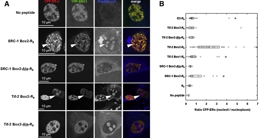
displace overexpressed full-length coactivators in living cells, Among the peptides tested, the highest ERa relocation score we tested the ERa binding of the synthetic molecular LXXLL was found for the SRC-1 Box2-R9, Tif-2 Box1-R9, and D2-R9 probes again, but now in the presence of the transfected SRC-1 peptides, with median ratios of ERa signal into the nucleoli and Tif-2 coactivators, tagged with yellow fluorescent protein versus the nucleoplasm of 1.2, 0.9, and 0.8, respectively (YFP). ERa indeed still colocalized with the nona-arginine (B). These cellular observations are in line with our peptides into the nucleoli except for the Tif-2 Box3- biochemical binding results B) and with previous reports R9, R9, and the alanine mutant peptides (We also indicating that ERa interacts with all three LXXLL motifs of the observed that the full-length SRC-1 coactivator could at least Figure 4. ERa-Binding Capacity of Syn-thetic Peptides in U2OS Cells in the Pres-ence of Overexpressed Coactivators(A) CLSM imaging of cells (positive events) trans-fected with ERa-CFP and SRC-1-YFP, in the pres-ence of estradiol (106 M). Scale bars represent10 mm.
(B) Quantification of ERa-nucleoli recruitment incell cotransfected with SRC-1-YFP expressed asthe ratio of the ERa-CFP fluorescence intensityper pixel into nucleoli versus the nucleoplasm.
Circles represent ratio values per cell (5 % n % 43).
Medians are indicated by the black line in themiddle of the box plots, whose sizes represent ±25% of the medians (program Profit). Asterisks (*)indicate results statistically different from thecontrol (*p < 0.01).
Chemistry & Biology 16, 702–711, July 31, 2009 ª2009 Elsevier Ltd All rights reserved 705


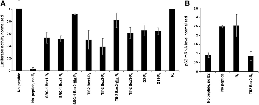
Chemistry & Biology Cellular Modulation of ERa with Synthetic Peptides Figure 5. Inhibition of ERa-Gene-MediatedTransactivation by Synthetic Nona-ArginineLXXLL Peptides(A) U2OS cells were transfected with ERa and theERa-DNA binding sequence ERE-tk-Luc. Lucif-erase activities are expressed as the ratio of Fireflyluciferase signal to Renilla luciferase signal and arenormalized to the ratio obtained when cells weresolely treated with the R9-tag peptide.
(B) Inhibition by R9 peptides of an endogenous ER-mediated pS2 gene in MCF-7 cells. MCF-7 cellswere cultured with 70 mM of the indicated peptidesin the presence of 10 nM estradiol (E2) (unless indi-cated). Cells were lysed after 3 hr of estradiol treat-ment, after which RNA was isolated and preparedfor QPCR analysis of endogenous ER-mediatedpS2 mRNA levels (normalized). Error bars repre-sent mean ± standard error.
partially be recruited to the nucleoli together with ERa in cells transcription (The largest inhibition, compared with treated with the most potent ERa-binding peptides (data not untreated cells, was obtained with the Tif-2 Box2-R9 peptide shown). This could be due to the recruitment of ERa as a dimer (61.0% of inhibition), the Tif-2 Box1-R9 peptide (49.8% of inhibi- to the nucleoli in which one monomer is bound to an arginine- tion), and the SRC-1 Box2-R9 peptide (48.2% of inhibition). For tagged peptide and the other monomer is still bound to SRC-1, comparison, when overexpressed in U2OS cells from a trans- or to the ligand-independent interaction between SRC-1 and fected DNA construct, an SRC-1 Box2 peptide exhibited a the AF-1 domain of ERa ( degree of inhibition (51.5%; similar to that of the Among the set of peptides tested, the strongest nucleoli synthetic SRC-1 Box2-R9 peptide on ERa-mediated gene tran- recruitment effect for ERa was observed with the Tif-2 Box2-R9 scription. The concentration dependence of the inhibition of peptide (median ratios, 2) and the SRC-1 Box2-R9 peptide the ERa transcriptional activity by the peptides was verified (median ratios, 0.7) with 95% and 88% of cells, with the Tif-2 peptide constructs (A), and no inhibition respectively, exhibiting a ratio higher than that found for cells was observed with external applied Tif-2 Box2 peptide without treated with the control peptide R9 ( an arginine tag B).
Similar experiments performed on cells cotransfected with We also examined the effect of the most efficacious peptide CFP-ERa and full-length YFP-Tif-2 coactivator followed by treat- from the luciferase reporter assay (Tif-2 Box2-R9) on the expres- ment with the synthetic SRC-1 Box2-R9 or Tif-2 Box2-R9 sion of an endogenous ERa target gene in the breast cancer cell peptides showed that these peptides could also efficiently line MCF-7 through quantitative polymerase chain reaction recruit ERa into the nucleoli in the presence of Tif-2 coactivator (QPCR) analysis MCF-7 cells treated with estradiol (It is interesting to note that ERa nucleoli-recruitment or with estradiol and the R9 peptide as reference showed the upon the Tif-2 Box2-R9 peptide treatment was found to be less expected up-regulation of ERa-mediated pS2 mRNA levels.
efficient when cells expressed the corresponding Tif-2 coactiva- MCF-7 cells treated with estradiol and the Tif-2 Box2-R9 peptide tor (median ratios, 0.6) than the SRC-1 coactivator (median featured pS2 mRNA levels analogous to cells not treated with ratios, 2), possibly indicating a higher affinity of the Tif-2 coacti- estradiol, showing the effective down-regulation of the pS2 vator or a different binding mechanism.
gene expression by the inhibitor peptide. Together, the resultsshow that inhibition of the ERa-coactivator interactions in cells Inhibition of ERa Gene Transactivation by exogenous synthetic peptides resulted in an inhibition of by Synthetic LXXLL Motif Molecular Probes ERa-mediated gene transcription, the extent of which depends The synthetic nona-arginine peptides induced sequestration of on the amino acid sequences of the peptides. There is thus ERa into the nucleoli. We therefore presumed a concomitant a clear relationship between the potential of the LXXLL molecular decrease of ERa transcriptional activity when perturbed with probes to bind and recruit ERa to the nucleoli and their capacity these peptides. Moreover, because these peptides are in fact to antagonize ERa-mediated gene transcription. The results mimicking the interactions between ERa and endogenous show that a cell-based study of the ERa binding and antago- cofactors, we expected that they might function in a dominant nizing activity of inhibitors directly evaluates their physiological negative manner when added to cells, disrupting these interac- tions and blocking the ERa transcriptional activity. To studysuch an activity, a firefly luciferase reporter gene controlled by EREs was cotransfected with an expression vector for ERa. Cellswere incubated in the presence or absence of estradiol, and the We have designed a set of synthetic peptidic LXXLL molecular reporter gene product was quantified in the resulting cell lysates probes bearing both a nona-arginine tag, promoting cell-entry by measuring the luminescence generated from the oxyluciferin and specific nucleoli localization, and a biotin tag, allowing the formed ). All the peptides, except for the control visualization of their intracellular localization. Neither the arginine R9 and alanine mutant peptides, decreased ERa gene-mediated tag nor the biotin epitope changed the binding affinity of the 706 Chemistry & Biology 16, 702–711, July 31, 2009 ª2009 Elsevier Ltd All rights reserved Chemistry & Biology Cellular Modulation of ERa with Synthetic Peptides peptides for the ERa LBD, showing that these tags are located at Tif-2 coactivators, normally found bound to ERa in the nucleo- sufficient distance from the binding pharmacophore. Even plasm. The probes thus can compete with full-length coactiva- though all the LXXLL-peptide sequences under study are tors in living cells. Quantification of ERa-nucleoli recruitment reported to have an affinity for the full-length ERa upon LXXLL-peptide treatment therefore provides, to our knowl- not all of the sequences were found edge, a new, simple, and efficient method to assess the potency to bind the ERa LBD strongly. The fact that the full-length of molecular probes to act as ERa-coactivator inhibitors under receptor can have a different surface topology as the isolated physiological conditions. Among the panel of peptides tested, the Tif-2 Box2-R9 and SRC-1 Box2-R9 peptides were found as ) is probably reflected in these observations, the most potent inhibitors of SRC-1 and Tif-2 coactivator binding.
especially since a cellular activity of all these LXXLL-peptide The specific physical inhibition of the ERa-coactivator interac- sequences could be shown in our studies.
tion could be correlated with a decrease in ERa transcriptional In order to perform subsequent cellular studies without the activity when perturbed with these peptides, both in a luciferase occurrence of any toxicity side effects, the concentrations at reporter assay and for activity of the endogenous pS2 gene which these probes showed cellular toxicity were determined.
in MCF-7 cells. Again, all LXXLL probes were functional and Interestingly, the peptides that featured both a nona-arginine decreased ERa gene-mediated luciferase transcription, whereas tag and an LXXLL-motif showed higher toxicity to the cells the control nona-arginine peptide and the AXXAA probes did not.
than either the nona-arginine tag alone or the peptide featuring In line with the nucleoli recruitment results, the Tif-2 Box2 and an arginine tag but with the LXXLL motif mutated to an AXXAA SRC-1 Box2 showed the strongest inhibition. The luciferase motif. Since no strong differences in cellular uptake are to be gene transcription is inhibited to around 50% This expected between the LXXLL and AXXAA peptides, the different value is also obtained when a similar peptide is overexpressed pharmacological behavior is more likely related to their intracel- in U2OS cells from a transfected DNA construct and in accor- lular effects. This furthermore indicates that the effects observed dance with literature data on overexpressed peptides ( for the molecular probes are highly sequence specific.
The Tif-2 Box2-R9 showed a CLSM imaging of cells incubated with these molecular probes full inhibition of the pS2 gene transcription in MCF-7 cells. The showed that they successfully entered the cells and adopted extent of the transcriptional inhibition may depend on the cell a particular localization into the nucleoli. This particular nucleoli type, assay format, and ligand-exposure time.
localization was not found for the non-R9-tagged peptides and It was previously shown that interaction of Tif-2 with ERa is is therefore exclusively due to the R9 tag. The efficient cellular strongly and selectively affected by mutations in NR-Box 2 uptake and specific nucleoli localization provide a strong meth- (). In a similar manner, it was shown that estra- odological tool to study any interactions of these molecular diol-bound full-length ERa displays unambiguous SRC-1 box probes with proteins, as it can be expected that either the local- preferences for NR-Box 2, while the recruitment of boxes 1 ization of the probes or that of the proteins will be perturbed upon and 3 of SRC-1 was not detected in vitro ().
binding. Transient transfection of cells with ERa together with The 75% homology found between the SRC-1 Box2 and Tif-2 their incubation with the LXXLL probes showed that these Box2 sequences provides a good explanation for the common peptides indeed resulted in perturbation of the ERa through features, both in terms of ERa-binding capacity and ERa-medi- recruitment to the nucleoli. This specific localization of ERa ated transcription inhibition that were observed in cells for these was not observed for a peptide consisting of the nona-arginine two peptides. Overall, there is a clear relationship between the tag alone, nor was it observed for the AXXAA-motif peptides or inhibitory effect on ERa-mediated gene transcription and ERa peptides not featuring the nona-arginine tag. The ERa recruit- translocation into the nucleoli by the molecular probes.
ment to the nucleoli by the nona-arginine LXXLL probes was The mechanism of ERa inhibition by ICBs typically is via furthermore found to be concentration dependent. These results a direct inhibition of the ERa-coativator interaction show that the observed effects are due to a specific interaction The nona-arginine-based inhibitors of the LXXLL motif with the ERa in combination with the localiza- reported here feature the same mechanism, but additionally tion properties of the nona-arginine tag. The fact that the nucleoli displace the ERa after inhibition of the ERa-coactivator interac- recruitment of ERa was observed for all the nona-arginine LXXLL tion. The mechanism of action is thus most likely a combination probes underlines the affinity of all these peptides for the full- of inhibition and sequestration of the receptor. The method of length ERa in a cellular context. The absence of this interaction cellular evaluation of inhibitory capacity on the full-length ERa in a biochemical context for some of these peptides on the iso- as presented here could therefore be applied to evaluate coac- lated ERa LBD shows that cellular evaluation, as presented tivator binding inhibitors in general. These inhibitors could be here, provides a more physiologically relevant view on the either provided themselves with localization and visualization binding interaction. The binding affinity of the peptides for the tags or be used in competition assays with the molecular peptide ERa can be evaluated by quantifying the ERa-nucleoli recruit- probes presented here. Also, this method can provide a basis to ment. This shows that certain peptide sequences, such as address NR selectivity for LXXLL-based inhibitors in a cellular SRC-1 Box2, Tif-2 Box1, and Tif-2 Box2, show a higher affinity context, when applied to a panel of different NRs.
than others. This is in line with observations that the ERa has The receptor's conformation is postulated to be a better indi- distinct preferences for the different NR boxes of coactivators cator of the antagonist activity of different ERa ligands than cell-based reporter gene assays ( The LXXLL probes maintain the property to recruit the ERa into ). The molecular probes presented the nucleoli even in the presence of overexpressed SRC-1 and here and derivatives thereof could provide a valuable entry to Chemistry & Biology 16, 702–711, July 31, 2009 ª2009 Elsevier Ltd All rights reserved 707 Chemistry & Biology Cellular Modulation of ERa with Synthetic Peptides diagnose the antagonist capacity of specific ligands in different The resin was washed 5 times with dichloromethane and ether successively cell types. In the presence of specific ERa antagonists, the and dried under vacuum for at least 1 hr. Cleavage of side chain protecting receptor adopts a conformation that prevents coactivators from groups and of the peptides from the resin was achieved by shaking the resinin a mixture of trifluoroacetic acid (TFA), triisopropyl silane, ethane-1,2-dithiol, binding ). It can be speculated that the molec- and H2O (95/1/2.5/1.5) for 2 hr. Then, the resin was removed by filtration and ular probes presented here will not interact with this antagonist- washed twice with TFA. Most of the TFA was evaporated from the combined liganded ERa. However, in the case of antagonist resistance, filtrates, and a 10-fold volume excess of cold diethyl ether was added drop- the ligand frequently acquires an agonistic function wise to achieve peptide precipitation. The crude peptides were isolated by and the molecular probes might centrifugation (3500 rpm, 10 min), washed with cold ether, taken up in water interact again with the ERa and thus induce nucleoli recruitment.
and lyophilized, and finally purified by reverse-phase high-performance liquidchromatography on a nucleodur C The method presented here will thus provide ways to probe the 18 Gravity column (125 3 21 mm, Macherey- Nagel) with a linear 60 min gradient of A (0.1% TFA in H2O) and B (0.1% TFA in surface of the ERa and determine its conformation in different MeCN) from 10% B to 100% B and a flow rate of 20 mL/min1. After purifica- cell-types, dependent on ligand, or with specific posttranslational tion, all the synthesized peptides were lyophilized and stored at 80C. The modifications or point mutations.
purity of the peptides was assessed by analytical liquid chromatography–mass spectrometry at 210 nm, 254 nm, and 280 nm using a diode array ultra-violet/visible detector. All the nona-arginine peptides were obtained pure with yields ranging from 13% to 41%.
The ERa-coactivator interaction, which is based on the In Vitro Competitive Fluorescence Polarization Assay Expression molecular recognition between consensus helical LXXLL and Purification of ERa-LBD Protein motifs of the coactivator and a hydrophobic pocket at the The pET15b-ERa-LBD plasmid (gift from Bayer-Schering Pharma AG) encod-ing the LBD of the human ERa was used as template for PCR amplification surface of the receptor, is an essential step for the ERa tran- using the primers 50-CCGGATGGATCCATGGGCAGCAGCCATCATCAT-30 scriptional activation process to take place. This interaction and 50-CCGGATCTCGAGTTAAGTGGGCGCATGTAGGCGG-30. The PCR is an important target for the development of anti–breast product (ERa-LBD residues 302–553) was digested with BamHI and XhoI cancers drugs. Here, a new, to our knowledge, chemical enzymes (Fermentas) and subcloned into a pHT401 plasmid to introduce a biology approach is reported in which synthetic LXXLL- Strep-tag and His-tag at the N terminus of the ERa-LBD construct. This peptide probes provided with specific visualization and plasmid (pHT503) was transformed into Escherichia coli Rosetta 2-DE3-placI localization tags interact directly with ERa in cells and per- (Novagen) cells that were then grown in Luria Bertani medium with 100 mg/Lampicilline and 34 mg/L chloramphenicol at 37C. At OD turb its localization and transcriptional activity. These 600nm 0.8, protein expression was induced with 0.5 mM isopropyl-beta-D-thiogalactopyranoside probes monitor the affinity of specific peptide sequences for 16–18 hr at 16C. Bacteria were then concentrated by centrifugation for for the full-length ERa in cells. This approach represents 10 min at 5,000 rpm and were kept at 80C until purification. Cells lysate a significant advancement over biochemical interaction chromatography was performed at 4C on an A¨KTA purifier using a HisTrap studies, which frequently focus on the isolated ERa LBD HP 5 ml column (GE Healthcare) pre-equilibrated with the protein extract alone. The molecular probes feature a nona-arginine tag buffer: 300 mM NaCl, 50 mM Na2HPO4, 2 mM NaH2PO4, and 20 mM imidazole(pH 8.0). Protein elution was obtained with 500 mM NaCl, 50 mM Na that facilitates cellular entry and also accounts for specific 2 mM NaH2PO4, 500 mM imidazole, and 10% (v/v) glycerol (pH 8.0); fractions subcompartmental localization at the nucleoli. This specific containing the ERa-LBD protein were then desalted on a Sephadex G25 feature allows these probes to dislocate the ERa from the PD-10 column (Amersham Biosciences) pre-equilibrated with 25 mM NaCl, DNA to the nucleoli upon binding and thus perturb the 20 mM Tris-HCl (pH 8.0), 10% (v/v) glycerol, and 1 mM tris (2-carboxyethyl) assembly of the receptor transcription complex. This molec- ular displacement of the ERa is accompanied by an inhibi-tion of its transcriptional activity. Quantification of the Fluorescence Polarization Measurements A reaction mixture containing a fluorescein-labeled coactivator (SRC-1 Box2) ERa-nucleoli recruitment is a simple and rapid method to peptide FL-CQLLTERHKILHRLLQEGSPSD (108 M), the ERa-LBD protein assess the affinity of peptide sequences for ERa in the phys- (106 M), and estradiol (105 M) was prepared. Fluorescence polarization inhi- iological context of a cell. Moreover, since the timing of addi- bition experiments were performed in 384-well plates (Optiplate-384 F, Perkin tion and concentration are easily controlled and their amino Elmer) by adding 10 ml of the reaction mixture to 40 ml of inhibitor peptides at acid sequences can be fully modified, these arginine-tagged increasing concentrations. After 2 hr of incubation at 4C, the fluorescence probes promise to be useful molecular tools to elucidate and polarization of the labeled coactivator peptide was measured on a plate readerfluorometer (Safire2TM, Tecan) with excitation at 470 nm and emission at modulate ERa functioning.
519 nm for fluorescein. The concentration of inhibitor peptide that resultedin a half-maximum decrease in the polarization value of the fluorescent coac- EXPERIMENTAL PROCEDURES tivator peptide displayed from the purified ERa was defined as IC50 and wasdetermined from three independent experiments with the ORIGIN 7 program Peptide Synthesis (Scientific Graphing and Analysis Software, OriginLab Corp).
The synthesis of all peptides was performed on solid support, using the Fmocstrategy. Biotin-PEG Nova Tag resin (0.48 mmol/g, Novabiochem) was Cell-Viability Assay allowed to swell in dimethylforamide (DMF) at room temperature for 30 min.
U2OS cancer cells (human osteosarcoma; ATCC number HTB-96) were split at After DMF was drained, the beads were incubated with 40% piperidine in 25% confluence in 96-well plates and maintained at 37C under 5% CO2 in DMF for the deprotection of the Fmoc group, and subsequently the amino Dulbecco's modified Eagle's medium (DMEM) supplemented with 1% (v/v) acids (AA) were introduced using the following coupling conditions: AA antibiotics penicillin/streptomycin (GIBCO) and 10% heat-inactivated fetal (3 eq), 2-(6-chloro-1H-benzotriazole-yl)-1,1,3,3-tetramethylaminium hexa- calf serum (GIBCO). After 1 day, cells were incubated with 100 ml of increasing fluorophosphate (3 eq), and N-methylmorpholine (6 eq) in DMF for 40 min.
concentrations of peptides (range from 0.01 to 5 g/L) directly diluted in the The Fmoc deprotection and introduction of amino acids was repeated until complete medium, for 14 hr. Subsequently, 10 ml of the cell proliferation the complete sequence was assembled.
reagent WST-1 was added to each well, and cells were incubated again in 708 Chemistry & Biology 16, 702–711, July 31, 2009 ª2009 Elsevier Ltd All rights reserved Chemistry & Biology Cellular Modulation of ERa with Synthetic Peptides a humidified atmosphere for 2.5 hr before measurement. The absorbance of cells were maintained at 37C under 5% CO2 for 4 hr before treatment with the samples was then measured using a microplate reader (Safire2TM, Tecan) synthetic peptides directly dissolved in the medium. For comparison, a at 450 nm, and cell toxicity was expressed as the percentage of inhibition of pcDNA3.1 plasmid encoding for the SRC-1 Box2 peptide sequence (pSF5 WST-1 incorporation, compared with control cells not treated with peptides.
vector) was also tested: cells were transfected with 20 ng of pSF5, 0.2 ng of All cell experiments were further performed at peptide concentrations that pCMV-HA-ERa, 200 ng of pERE-tK-firefly luciferase, and 2 ng of SV40 Renilla did not induce more than 20% of cell toxicity.
luciferase plasmids. Four hours after adding the peptides, estradiol (108 M)was incubated with the cells for 16 hr before the reading of firefly luciferase Cell Imaging by Confocal Laser Scanning Microscopy (CLSM): expression by a dual-luciferase reporter assay system (Promega). Lumines- Cellular Localization of the Synthetic Peptides cent intensities were recorded on a Berthold Centro XS3 LB 960 (Berthold U2OS cells were cultured on coverslips for 48 hr before imaging in the same technologies). Firefly luciferase activity signal was first normalized with respect complete medium as mentioned above. Fourteen hours before imaging, cells to the constitutive R. reniformis luciferase signal and second normalized to the were incubated with peptides at the nontoxic peptide concentrations luciferase activity found in control cells incubated with the R9 tag alone.
described above and then were fixed with 4% paraformaldehyde in PBS for Results shown are representative of three independent experiments.
10 min at room temperature. A 10-min treatment of the fixed cells by PBS-NH4Cl 50 mM and then PBS-Glycine 20 mM was used to avoid self-fluores- pS2 Gene Inhibition in MCF-7 Cells cence bleaching. Cells were permeabilized with Triton X-100 0.1% for For quantitative RT-PCR in MCF-7 cells, cells were cultured in phenol red–free 10 min, washed 3 times, and incubated for 30 min with a blocking solution DMEM containing 5% charcoal-treated serum for 72 hr prior to the onset of the of PBS-BSA 0.1%. The cellular distribution of peptides was revealed after experiment in order to deplete activated ER in the cells. Subsequently, cells immunolabeling of the cells using a Cy5-conjugated monoclonal mouse anti- were cultured overnight in the presence of the indicated peptides at a final biotin antibody (Jackson ImmunoResearch) diluted 1:400 in PBS and their concentration of 70 mM. After overnight treatment, 10 nM estradiol was added CLSM imaging (Leica TCS SP2), with the following settings: lex = 633 nm for 3 hr or the cells were left untreated. After exposure to hormones, cells were and lem = 650–700 nm.
lysed, and RNA was extracted using Trizol (Invitrogen), according to the manu-facturer's protocol. RNA was reverse-transcribed using SuperScript(tm) III Cellular Assessment of ERa-LXXLL-Peptides Binding Reverse Transcriptase (Invitrogen), on which QPCR was performed using U2OS cells were cultured on coverslips in a 6-well plate for 48 hr before CYBR Green (Applied Biosystems), according to the manufacturer's proto- imaging in DMEM without phenol red supplemented with 1% penicillin/strep- cols. The pS2 cDNA was amplified with the forward primer 50 CATCGAC tomycin and 10% hormone-depleted (charcoal/dextran treated) and heat- GTCCCTCCAGAAGA and the reverse primer 50 CTCTGGGACTAATCACCG inactivated fetal calf serum (Thermo Scientific HyClone). At 50% of confluence, TGCT. As a control for equal loading, the observed signals were related to U2OS cells (neither expressing ERa nor ERb) were tran- b-actin RNA levels, using a forward primer 50 CCTGGCACCCAGCACAAT siently transfected by PEI (Polysciences) or Fugene 6 (Roche) with 1 mg of and reverse primer 50 GGGCCGGACTCGTCATACT. Input levels were related pcDNA3.1 plasmid containing the HA-ERa-CFP construct to b-actin house keeping gene mRNA levels. Average values and SD were Cells were incubated overnight with the nona-arginine peptides dissolved in obtained for samples without peptide (duplicate) or with peptide (triplicate).
the culturing medium. Afterward, estradiol was added to the cultures at a finalconcentration of 106 M for 3 hr. Cells were then fixed and stained asdescribed above and imaged by CLSM (objective X63) with the followed SUPPLEMENTAL DATA independent settings: lex = 458 nm and lem = 470–500 nm for CFP, andlex = 633 nm and lem = 650–700 nm for Cy5.
Supplemental data include two tables and five figures and can be found with The quantification of ERa-CFP in the nucleoli was performed with the Quan- this article online at tify Histogram module within the CLSM Leica software. First, on the basis of Cy5 fluorescence, for each cell, regions of interest (ROIs) were designed,encompassing the nucleoli containing peptides and the nucleoplasm. Then, the average CFP fluorescence intensity per pixel in the ROIs were subtractedby the fluorescence intensity of the background to calculate the enrichment of We thank Emmanuel Margeat and Julien Savatier (Centre de Biochimie ERa-CFP in the nucleoli compared to the nucleoplasm as the mean ratio (CFP Structurale, Montpellier, France) for advice on microscopy and for providing intensity nucleoli/CFP intensity nucleoplasm). Results are presented as the the pcDNA3-YFP-Tif-2 plasmid. We thank Sascha Fuchs (Chemical Genomic ratios (nucleoli/nucleoplasm) found per peptide for two independent experi- Centre, Dortmund, Germany) for providing the pSF5 plasmid. We thank Scher- ments, for a 10–20 cell population.
ing Plough, Bayer-Schering Pharma, and Merck-Serono for financial support.
W. Zwart was supported by TI Pharma project T3-107.
Cellular Assessment of LXXLL Peptides/Coactivators (SRC-1 and Tif-2) Competitive Binding for ERa Received: February 6, 2009 U2OS cells were cultivated and treated in the same conditions as above and Revised: May 21, 2009 transiently cotransfected with 1 mg of pcDNA3-ERa-CFP plasmid and 2 mg Accepted: June 19, 2009 of pcDNA3-SRC-1-YFP plasmid or 2 mg of pcDNA3-YFP-Tif-2 plasmid. After Published: July 30, 2009 cells were fixed and labeled with the Cy5-conjugated anti-biotin antibody,they were imaged by CLSM with the successive settings of CFP, Cy5, and YFP (lex = 514 nm and lem = 525–550 nm). The same method as describedabove was used to quantify ERa-CFP fluorescence into the nucleoli and was Bapat, A.R., and Frail, D.E. (2003). Full-length estrogen receptor alpha and its compared to the one found in the cytoplasm.
ligand-binding domain adopt different conformations upon binding ligand.
J. Steroid Biochem. Mol. Biol. 86, 143–149.
Inhibition of ERa-Mediated Gene Transcription ERa Luciferase Bedner, E., Li, X., Gorczyca, W., Melamed, M.R., and Darzynkiewicz, Z. (1999).
Transactivation Assay Analysis of apoptosis by laser scanning cytometry. Cytometry 35, 181–195.
U2OS cells were split in 24-well plates and maintained at 37C under 5% CO2in Dulbecco's modified Eagle's medium (DMEM) without phenol red, supple- Berridge, M.V., Herst, P.M., and Tan, A.S. (2005). Tetrazolium dyes as tools in mented with 1% (v/v) antibiotics penicillin/streptomycin and 10% hormone- cell biology: new insights into their cellular reduction. Biotechnol. Annu. Rev.
depleted and heat-inactivated fetal calf serum (Thermo Scientific HyClone).
After 1 day, cells were transfected using PEI with 10 ng of pCMV-HA-ERa Bindels, E.M., Lallemand, F., Balkenende, A., Verwoerd, D., and Michalides, R.
full length, 200 ng of pERE-tK-firefly luciferase (), and (2002). Involvement of G1/S cyclins in estrogen-independent proliferation of 2 ng of SV40 Renilla reniformis luciferase plasmids. After DNA transfection, estrogen receptor-positive breast cancer cells. Oncogene 21, 8158–8165.
Chemistry & Biology 16, 702–711, July 31, 2009 ª2009 Elsevier Ltd All rights reserved 709 Chemistry & Biology Cellular Modulation of ERa with Synthetic Peptides Bramlett, K.S., Wu, Y., and Burris, T.P. (2001). Ligands specify coactivator Kurebayashi, S., Nakajima, T., Kim, S.C., Chang, C.Y., McDonnell, D.P., nuclear receptor (NR) box affinity for estrogen receptor subtypes. Mol. Endo- Renaud, J.P., and Jetten, A.M. (2004). Selective LXXLL peptides antagonize crinol. 15, 909–922.
transcriptional activation by the retinoid-related orphan receptor RORgamma.
Chang, C., Norris, J.D., Grøn, H., Paige, L.A., Hamilton, P.T., Kenan, D.J., Biochem. Biophys. Res. Commun. 315, 919–927.
Fowlkes, D., and McDonnell, D.P. (1999). Dissection of the LXXLL nuclear LaFrate, A.L., Gunther, J.R., Carlson, K.E., and Katzenellenbogen, J.A. (2008).
receptor-coactivator interaction motif using combinatorial peptide libraries: Synthesis and biological evaluation of guanylhydrazone coactivator binding discovery of peptide antagonists of estrogen receptors alpha and beta. Mol.
inhibitors for the estrogen receptor. Bioorg. Med. Chem. 16, 10075–10084.
Cell. Biol. 19, 8226–8239.
Larson, C.J., Osburn, D.L., Schmitz, K., Giampa, L., Mong, S.M., Marschke, K., Chang, C.Y., and McDonnell, D.P. (2002). Evaluation of ligand-dependent Seidel, H.M., Rosen, J., and Negro-Vilar, A. (2005). Peptide binding identifies changes in AR structure using peptide probes. Mol. Endocrinol. 16, 647–660.
an ERalpha conformation that generates selective activity in multiple in vitro Chang, C.Y., Abdo, J., Hartney, T., and McDonnell, D.P. (2005). Development assays. J. Biomol. Screen. 10, 590–598.
of peptide antagonists for the androgen receptor using combinatorial peptide Leduc, A.M., Trent, J.O., Wittliff, J.L., Bramlett, K.S., Briggs, S.L., Chirgadze, phage display. Mol. Endocrinol. 19, 2478–2490.
N.Y., Wang, Y., Burris, T.P., and Spatola, A.F. (2003). Helix-stabilized cyclic Clarke, R., Leonessa, F., Welch, J.N., and Skaar, T.C. (2001). Cellular and peptides as selective inhibitors of steroid receptor-coactivator interactions.
molecular pharmacology of antiestrogen action and resistance. Pharmacol.
Proc. Natl. Acad. Sci. USA 100, 11273–11278.
Rev. 53, 25–71.
Mahajan, M.A., and Samuels, H.H. (2005). Nuclear hormone receptor coregu- Connor, C.E., Norris, J.D., Broadwater, G., Willson, T.M., Gottardis, M.M., lator: role in hormone action, metabolism, growth, and development. Endocr.
Dewhirst, M.W., and McDonnell, D.P. (2001). Circumventing tamoxifen resis- Rev. 26, 583–597.
tance in breast cancers using antiestrogens that induce unique conformational McInerney, E.M., Rose, D.W., Flynn, S.E., Westin, S., Mullen, T.M., Krones, A., changes in the estrogen receptor. Cancer Res. 61, 2917–2922.
Inostroza, J., Torchia, J., Nolte, R.T., Assa-Munt, N., et al. (1998). Determinants Ding, X.F., Anderson, C.M., Ma, H., Hong, H., Uht, R.M., Kushner, P.J., and of coactivator LXXLL motif specificity in nuclear receptor transcriptional Stallcup, M.R. (1998). Nuclear receptor-binding sites of coactivators glucocor- activation. Genes Dev. 12, 3357–3368.
ticoid receptor interacting protein 1 (GRIP1) and steroid receptor coactivator 1 McKenna, N.J., Lanz, R.B., and O'Malley, B.W. (1999). Nuclear receptor (SRC-1): multiple motifs with different binding specificities. Mol. Endocrinol.
coregulators: cellular and molecular biology. Endocr. Rev. 20, 321–344.
Michalides, R., Griekspoor, A., Balkenende, A., Verwoerd, D., Janssen, L., Fisher, B., Costantino, J.P., Wickerham, D.L., Cecchini, R.S., Cronin, W.M., Jalink, K., Floore, A., Velds, A., van't Veer, L., and Neefjes, J. (2004). Tamoxifen Robidoux, A., Bevers, T.B., Kavanah, M.T., Atkins, J.N., Margolese, R.G., resistance by a conformational arrest of the estrogen receptor alpha after PKA et al. (2005). Tamoxifene for the prevention of breast cancer: current status activation in breast cancer. Cancer Cell 5, 597–605.
of National Surgical Adjuvant Breast and Bowel Project P-1 study. J. Natl.
Monroe, D.G., Getz, B.J., Johnsen, S.A., Riggs, B.L., Khosla, S., and Cancer Inst. 97, 1652–1662.
Spelsberg, T.C. (2003). Estrogen receptor isoform-specific regulation of Futaki, S., Suzuki, T., Ohashi, W., Yagami, T., Tanaka, S., Ueda, K., and endogenous gene expression in human osteoblastic cell lines expressing either ERalpha or ERbeta. J. Cell. Biochem. 90, 315–326.
membrane-permeable peptides having potential as carriers for intracellular Norris, J.D., Paige, L.A., Christensen, D.J., Chang, C.Y., Huacani, M.R., Fan, protein delivery. J. Biol. Chem. 276, 5836–5840.
D., Hamilton, P.T., Fowlkes, D.M., and McDonnell, D.P. (1999). Peptide Galande, A.K., Bramlett, K.S., Burris, T.P., Wittliff, J.L., and Spatola, A.F.
antagonists of the human estrogen receptor. Science 285, 744–746.
(2004). Thioether side chain cyclization for helical peptide formation: inhibitors Osborne, C.K., Bardou, V., Hopp, T.A., Chamness, G.C., Hilsenbeck, S.G., of estrogen receptor-coactivator interactions. J. Pept. Res. 63, 297–302.
Fuqua, S.A., Wong, J., Allred, D.C., Clark, G.M., and Schiff, R. (2003). Role Galande, A.K., Bramlett, K.S., Trent, J.O., Burris, T.P., Wittliff, J.L., and of the estrogen receptor coactivator AIB1 (SRC-3) and HER-2/neu in tamoxifen Spatola, A.F. (2005). Potent inhibitors of LXXLL-based protein-protein interac- resistance in breast cancer. J. Natl. Cancer Inst. 95, 353–361.
tions. Chem. Bio. Chem. 6, 1991–1998.
Parent, A.A., Gunther, J.R., and Katzenellenbogen, J.A. (2008). Blocking Geistlinger, T.R., McReynolds, A.C., and Guy, R.K. (2004). Ligand-selective estrogen signaling after the hormone: pyrimidine-core inhibitors of estrogen inhibition of the interaction of steroid receptor coactivators and estrogen receptor-coactivator binding. J. Med. Chem. 51, 6512–6530.
receptor isoforms. Chem. Biol. 11, 273–281.
Pike, A.C., Brzozowski, A.M., and Hubbard, R.E. (2000). A structural biologist's Girault, I., Lerebours, F., Amarir, S., Tozlu, S., Tubiana-Hulin, M., Lidereau, R., view of the oestrogen receptor. J. Steroid Biochem. Mol. Biol. 74, 261–268.
and Bie che, I. (2003). Expression analysis of estrogen receptor alpha coregu- Rishi, A.K., Shao, Z.M., Baumann, R.G., Li, X.S., Sheikh, M.S., Kimura, S., lators in breast carcinoma: evidence that NCOR1 expression is predictive of Bashirelahi, N., and Fontana, J.A. (1995). Estradiol regulation of the human the response to tamoxifen. Clin. Cancer Res. 9, 1259–1266.
retinoic acid receptor alpha gene in human breast carcinoma cells is mediated Gunther, J.R., Moore, T.W., Collins, M.L., and Katzenellenbogen, J.A. (2008).
via an imperfect half-palindromic estrogen response element and Sp1 motifs.
Amphipathic benzenes are designed inhibitors of the estrogen receptor alpha/ Cancer Res. 55, 4999–5006.
steroid receptor coactivator interaction. ACS Chem. Biol. 3, 282–286.
Rodriguez, A.L., Tamrazi, A., Collins, M.L., and Katzenellenbogen, J.A. (2004).
Hall, J.M., Chang, C.Y., and McDonnell, D.P. (2000). Development of peptide Design, synthesis, and in vitro biological evaluation of small molecule inhibitors antagonists that target estrogen receptor beta-coactivator interactions.
of estrogen receptor alpha coactivator binding. J. Med. Chem. 47, 600–611.
Mol. Endocrinol. 14, 2010–2023.
Schaufele, F., Chang, C.Y., Liu, W., Baxter, J.D., Nordeen, S.K., Wan, Y., Day, Holst, F., Stahl, P.R., Ruiz, C., Hellwinkel, O., Jehan, Z., Wendland, M., R.N., and McDonnell, D.P. (2000). Temporally distinct and ligand-specific Lebeau, A., Terracciano, L., Al-Kuraya, K., Ja¨nicke, F., et al. (2007). Estrogen recruitment of nuclear receptor-interacting peptides and cofactors to subnu- receptor alpha (ESR1) gene amplification is frequent in breast cancer. Nat.
clear domains containing the estrogen receptor. Mol. Endocrinol. 14, 2024– Genet. 39, 655–660.
Howell, A., DeFriend, D., Robertson, J., Blamey, R., and Walton, P. (1995).
Shao, D., Berrodin, T.J., Manas, E., Hauze, D., Powers, R., Bapat, A., Gonder, Response to a specific antioestrogen (ICI 182780) in tamoxifen-resistant D., Winneker, R.C., and Frail, D.E. (2004). Identification of novel estrogen breast cancer. Lancet 345, 29–30.
receptor alpha antagonists. J. Steroid Biochem. Mol. Biol. 88, 351–360.
Kosuge, M., Takeuchi, T., Nakase, I., Jones, A.T., and Futaki, S. (2008). Cellular Shao, G., Heyman, R.A., and Schulman, I.G. (2000). Three amino acids specify internalization and distribution of arginine-rich peptides as a function of extra- coactivator choice by retinoid X receptors. Mol. Endocrinol. 14, 1198–1209.
cellular peptide concentration, serum, and plasma membrane associated Shiau, A.K., Barstad, D., Loria, P.M., Cheng, L., Kushner, P.J., Agard, D.A., and proteoglycans. Bioconjug. Chem. 19, 656–664.
Greene, G.L. (1998). The structural basis of estrogen receptor/coactivator 710 Chemistry & Biology 16, 702–711, July 31, 2009 ª2009 Elsevier Ltd All rights reserved Chemistry & Biology Cellular Modulation of ERa with Synthetic Peptides recognition and the antagonism of this interaction by tamoxifen. Cell 95, Webb, P., Nguyen, P., Shinsako, J., Anderson, C., Feng, W., Nguyen, M.P., Chen, D., Huang, S.M., Subramanian, S., McKinerney, E., et al. (1998).
Stenoien, D.L., Mancini, M.G., Patel, K., Allegretto, E.A., Smith, C.L., and Estrogen receptor activation function 1 works by binding p160 coactivator Mancini, M.A. (2000). Subnuclear trafficking of estrogen receptor-alpha and proteins. Mol. Endocrinol. 12, 1605–1618.
steroid receptor coactivator-1. Mol. Endocrinol. 14, 518–534.
Xu, J., Qiu, Y., DeMayo, F.J., Tsai, S.Y., Tsai, M.J., and O'Malley, B.W. (1998).
Tu¨nnemann, G., Martin, R.M., Haupt, S., Patsch, C., Edenhofer, F., and Partial hormone resistance in mice with disruption of the steroid receptor co- Cardoso, M.C. (2006). Cargo-dependent mode of uptake and bioavailability activator-1 (SRC-1) gene. Science 279, 1922–1925.
of TAT-containing proteins and peptides in living cells. FASEB J. 20, 1775– Xu, J., Liao, L., Ning, G., Yoshida-Komiya, H., Deng, C., and O'Malley, B.W.
(2000). The steroid receptor coactivator SRC-3 (p/CIP/RAC3/AIB1/ACTR/ Tu¨nnemann, G., Ter-Avetisyan, G., Martin, R.M., Sto¨ckl, M., Herrmann, A., and TRAM-1) is required for normal growth, puberty, female reproductive function, Cardoso, M.C. (2008). Live-cell analysis of cell penetration ability and toxicity and mammary gland development. Proc. Natl. Acad. Sci. USA 97, 6379–6384.
of oligo-arginines. J. Pept. Sci. 14, 469–476.
Zwart, W., Griekspoor, A., Berno, V., Lakeman, K., Jalink, K., Mancini, M., Vaz, B., Mo¨cklinghoff, S., and Brunsveld, L. (2008). Targeting the nuclear- Neefjes, J., and Michalides, R. (2007a). PKA-induced resistance to tamoxifen cofactor interaction. In Nuclear receptors as drug targets, E. Ottow and H.
is associated with an altered orientation of ERalpha towards co-activator SRC- Weinmann, eds. (Weinheim, Germany: Wiley-VCH Verlag GmbH), pp. 25–40.
1. EMBO J. 26, 3534–3544.
Voegel, J.J., Heine, M.J., Tini, M., Vivat, V., Chambon, P., and Gronemeyer, H.
Zwart, W., Griekspoor, A., Rondaij, M., Verwoerd, D., Neefjes, J., and (1998). The coactivator TIF2 contains three nuclear receptor-binding motifs Michalides, R. (2007b). Classification of anti-estrogens according to intramo- and mediates transactivation through CBP binding-dependent and -indepen- lecular FRET effects on phospho-mutants of estrogen receptor alpha. Mol.
dent pathways. EMBO J. 17, 507–519.
Cancer Ther. 6, 1526–1533.
Chemistry & Biology 16, 702–711, July 31, 2009 ª2009 Elsevier Ltd All rights reserved 711

Source: http://mate.tue.nl/mate/pdfs/11287.pdf

Untitled

BioFactors 25 (2005) 97–107 Coenzyme Q10 protects SHSY5Y neuronalcells from beta amyloid toxicity andoxygen-glucose deprivation by inhibiting theopening of the mitochondrial permeabilitytransition pore Geng Lia,∗, Liang-Yu Zoua,b, Chun-Mei Caoc and Edward S. Yanga aThe Jockey Club MRI Centre, The University of Hong Kong, Hong Kong, China bDepartment of Neurology, The First Affiliated Hospital, Harbin Medical University, China

7th kelantan health conference 15 –16th june 201

Volume 11(Suppl 3) 2011 Official Journal of Malaysian P ublic Health Physicians'Association EDITORIAL BOARD Chief Editor Prof. Dato' Dr. Syed Mohamed Aljunid (Unit ed Nations University – International Institute for Global Health) Deputy Chief Editor Assc. Prof. Dr. Sharifa Ezat Wan Puteh