Mff circular 39
Entomology Circular No. 406
Fla. Dept. Agric. & Consumer Services
Division of Plant Industry
The Present Status and a Review of the Brown Recluse and Related
Spiders, Loxosceles spp. (Araneae: Sicariidae), in Florida1
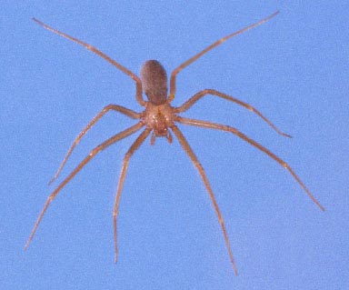
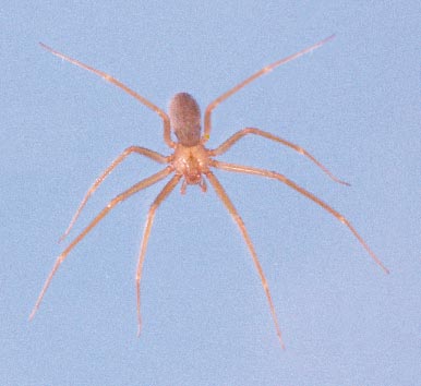
INTRODUCTION: The brown recluse spider, Loxosceles reclusa Gertsch & Mulaik (Figs. 1-2, 6), is frequently
reported in Florida as a cause of necrotic lesions in humans. For example, in the year 2000 alone, Loft (2001) reported
that the Florida Poison Control Network had recorded nearly 300 alleged cases of brown recluse bites in the state; a
subset of 95 of these bites was reported in the 21 counties (essentially central Florida) under the jurisdiction of the
regional poison control center in Tampa. I called the Florida Poison Control Network to confirm these numbers, and was
cited 182 total cases and 96 in the Tampa region. The actual numbers are less important than the fact that a significant
number of unconfirmed brown recluse spider bites are reported in the state every year. Yet not one specimen of brown
recluse spider has ever been collected in Tampa, and the only records of Loxosceles species in the entire region are from
Orlando and vicinity. A general review of the brown recluse, along with a critical examination of the known distribution
of brown recluse and related spiders in Florida, seems in order at this time.
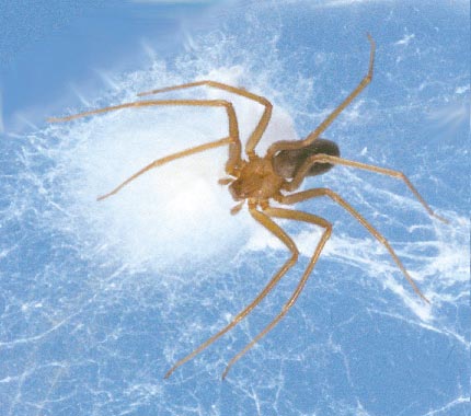
Fig. 1. Female brown recluse spider (2X natural size); 2. Female with eggsac. Photography credits: Jeffrey Lotz.
DISTRIBUTION: Loxosceles reclusa was described by Gertsch and Mulaik (1940) from Texas. At the time of the first
revision of the genus Loxosceles in the Americas (Gertsch 1958), the known distribution ranged from Central Texas to
southern Kansas, east through middle Missouri to western Tennessee and northern Alabama, and south to southern
Mississippi. Gorham (1968) added Illinois, Kentucky and northern Georgia. Later, he added Nebraska, Iowa, Indiana
and Ohio, with scattered introductions in other states, including Florida; his map indicated a record in the vicinity of
Tallahassee (Gorham 1970). Weems and Whitcomb (1975) noted that, "on many occasions specimens have been
inadvertently brought into Florida in trucks and automobiles, hidden in luggage, boxes, and various commercial cargoes,
but to date it appears to have been unsuccessful in establishing breeding populations in Florida." It is unfortunate that
they did not document these alleged records, as this comment is not completely in accord with the following reference.
An updated revision of the genus by Gertsch and Ennik (1983) reported a few records from Arizona, California,
Colorado, Florida, Maine, Minnesota, New Jersey, New Mexico, New York, North Carolina, Wyoming and Tamaulipas
(Mexico) [the reported Ontario (Canada) record in this publication subsequently proved to be a specimen of Loxosceles
rufescens (Dufour); R. Vetter, pers. comm., 2001]. Most of these peripheral records were interceptions of one or two
1Entomology contribution No. 918; Bureau of Entomology, Nematology & Plant Pathology - Entomology Section.
2Curator: Arachnida & Myriapoda, Florida State Collection of Arthropods, FDACS, Division of Plant Industry, P. O. Box 147100, Gainesville, FL 32614-7100.
specimens, not evidence of established populations. The Florida records consisted of two specimens, one each from
Alachua (collected 10 January 1969) and Jefferson (Monticello, collected 21 August 1968) counties, and both were taken
from inside automobiles. Subsequently, a sailor was bitten on the hand by a male brown recluse in the cargo hold of a
naval ship in Jacksonville, in March 1986. This ship had just arrived from North Carolina, where it had loaded supplies.
To date, this appears to be the only verified case (the actual causative agent of a bite captured and identified) of brown
recluse spider bite in Florida [due to complicating factors, medical personnel familiar with this case even questioned the
veracity of this one alleged bite].
Within the last two years, single buildings (in Callaway, Jacksonville and Tallahassee) have been found to contain
populations of L. reclusa (Edwards 1999, 2000, 2001). There is reason to believe that all three of these records are theresult of movement of infested materials from other states, so it is entirely possible that the infestations are restricted tothese buildings and can be eliminated. Such was the case with an infestation of the similar L. rufescens found in OrangeCounty (DPI records from Orlando: 28 January 1982, 4 January 1983 and 18 August 1986) in a single building; thespiders were subsequently eradicated. The only other records of L. rufescens occurring in Florida are a few juvenilespiders in buildings in nearby Osceola County (Runnymede; Banks 1904) and one juvenile specimen from Dade County(Lemon City; Gertsch 1958). This cosmopolitan species is probably native to the Mediterranean region, and issometimes called the Mediterranean brown spider or Mediterranean recluse. It has been recorded from a number oflocalities across the U.S., particularly in larger cities, where it is transported by commerce (Gertsch and Ennik 1983). Noother species of Loxosceles has been reported from Florida.
In summary, the verified records of brown recluse and related spiders in the state are limited to the following eight
out of 67 Florida counties: Alachua, Bay (Callaway), Dade (Lemon City), Duval (Jacksonville), Jefferson (Monticello),Leon (Tallahassee), Orange (Orlando) and Osceola (Runnymede). The more northern counties (Alachua, Bay, Duval,Jefferson and Leon) were all isolated records of the native brown recluse, L. reclusa, whereas the more southern countyrecords (Dade, Orange and Osceola) were of the introduced Mediterranean recluse, L. rufescens. The Alachua, Dade andJefferson county records were interceptions of single specimens. The Bay, Duval, Leon, Orange and Osceola countyrecords were infestations in one or two buildings. There is no evidence to support either the notion that a widespreadpopulation of brown recluse spiders exists in Florida or that there are numerous introductions of brown recluse into thestate. Therefore, there is no reason to assume that frequent interactions between brown recluse and humans occur inFlorida.
I have personally identified several hundred Florida spiders submitted for identification by the public, and only one
specimen (the Bay County record) proved to be a brown recluse spider. In addition, I have seen thousands of Floridaspiders submitted by professional biologists and inspectors, with only the few specimens mentioned above proving to bemembers of the genus Loxosceles. It appears obvious to me that the chance of interaction between brown recluse spidersand people in Florida is close to nil, agreeing with Vetter's (2000) assessment of reported brown recluse bites outside thenatural range of the spider. Medical personnel should, therefore, consider a multitude of more likely causes (see below)before diagnosing and treating a necrotic wound as a brown recluse bite.
DESCRIPTION: The description is taken from Gertsch (1958). Adults of both sexes are similar in appearance and size,
ranging from about 7-12 mm in body length. Adult females average slightly larger, about 9 mm compared to about 8 mm
for adult males. The carapace is pale yellow to reddish brown, with a dusky brown patch just in front of the median
groove (which is encompassed by a narrow, dark line); this patch is united to the front of the carapace by dusky brown
stripes. In total, these markings appear in the form of a violin. In addition, three dusky patches may occur along the
margin on each side. The sternum is yellowish, with other ventral body parts of the cephalothorax darker reddish brown.
The legs are slender and dusky orange to dark reddish brown. They are numbered front to back with Roman numerals (I,
II, III, IV). In females, the leg length formula, longest to shortest, is II, IV, I, III, typically with leg II being over 18 mm
in length, and leg III about 15 mm, the other two pair intermediate in length. The male leg formula is II, I, IV, III, with
leg II over 24 mm and leg III about 17 mm. The abdomen of both sexes is tan to brown, but it may appear darker if the
spider has recently fed. Juveniles are paler in all respects, as are occasional adults.
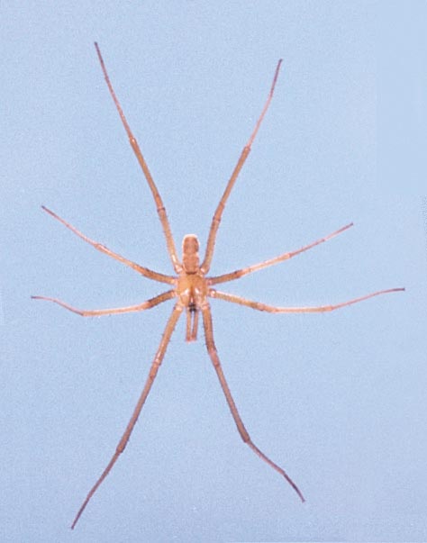
Males of the common southern crevice spider (Fig. 3), Kukulcania (formerly Filistata) hibernalis (Hentz), are
frequently confused with the brown recluse (Edwards 1983). The male palp length of L. reclusa is under 4 mm,considerably less than the superficially similar crevice spider. Another difference between the two species is that L.
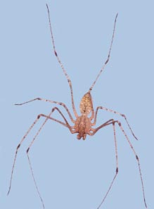
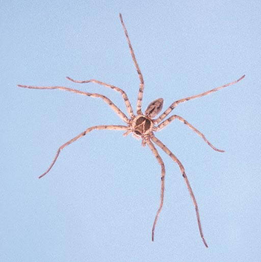
reclusa has six eyes composed of three isolated pairs (dyads), whereas K. hibernalis has eight eyes all clumped togetherin the middle front of the carapace. The only other Florida spiders with an eye arrangement similar to Loxosceles are thespitting spiders of the genus Scytodes (Fig. 4), but these spiders have a domed carapace, lack a violin-shaped carapacemarking, and are not known to cause serious wounds in humans. Occasionally, the huntsman spider (Fig. 5), Heteropodavenatoria (L.), is misidentified as a brown recluse (Edwards 1979). However, the color pattern on the carapace of thisspecies is reversed, with a light median mark on a dark background, and adults of this spider are much larger than abrown recluse.
Entomology Circular No. 406
BIOLOGY: Hite et al. (1966) made the earliest thorough report of the biology of L. reclusa. In their study, they
recorded the habitat of 626 brown recluse in Arkansas found from May 1962 to December 1964. Most (430) were found
in buildings and outbuildings, especially in boxes and among papers, in every room from basement to attic. They were
found in almost any place which had remained undisturbed for lengthy periods of time, such as behind pictures, beneath
or behind furniture, in boxes of toys, in clothing, among stored papers, in the corrugations of cardboard boxes, and in
discarded articles, such as tires, inner tubes, and assorted other junk. Most of the specimens found in feral conditions
were under rocks, especially in bluff outcrops, with a few under bark or in logs. They definitely seemed to prefer dry
conditions.
Spiderlings appear to stay with their mother for 3-4 instars before dispersing. They feed on prey provided by the
mother during this time. Once dispersed, they may establish a home territory, where they stay through several moreinstars, as evidenced by the presence of several successive molts. Spiders go through a total of eight instars. Irregularwebbing is seen in the nest area. Prey consists of a variety of other arthropods, including rather dangerous prey like otherspiders and ants. The attack consists of a sudden lunge and bite, usually on an intended prey's appendage, after which thebrown recluse immediately backs away (personal observation). The venom acts rapidly to paralyze the prey, preventingany retaliation for the initial attack of the recluse spider. After the prey is overcome by the venom, the brown reclusemoves in to feed. Relatively harmless prey, particularly mobile prey like houseflies, will be held with the initial bite andnot released.
Most mating and reproduction occurs during June and July. Females were frequently found with more than one
eggsac (Fig. 2). In the laboratory, females made up to five eggsacs. Total eggs per female ranged from 31-300, totalhatched young maximized at 158 for a single female; the largest number of young from one eggsac was 91; and percentemergence of young was 0-100. Some eggs were fed on by spiderlings from previous eggsacs still in the web, or even bythe female (perhaps these were infertile). The egg stage averaged about 13 days, instars I-VIII 17, 110, 63, 41, 38, 34,40, and 53 days respectively. Maximum age for a brown recluse from emergence to death was 894 days for a female, 796for a male. A laboratory-kept specimen lived over six months without food or water. Captive specimens also provedmoderately resistant to pesticides. These two characteristics illustrate why brown recluse populations may exist inbuildings for long periods of time, despite repeated efforts to eradicate them.
BITES AND BITE SYMPTOMS: Brown recluse spiders usually bite only when they become trapped next to the
victim's skin. Bites occur either when sleeping humans roll onto the spider or put on clothes into which the spider has
crawled (Vetter and Visscher 1998). Typically bites occur under clothing, mostly on the thigh, upper arm, or lateral
torso, less often on the neck (Anderson 1998) [Dr. Philip C. Anderson is a physician and medical researcher who has
worked on brown recluse bites and venom for 40 years].
Description of the symptoms is from Wingo (1960), Gorham (1968, 1970), Anderson (1982, 1998), and Vetter and
Visscher (1998). Reactions to a bite vary from no noteworthy symptoms to severe necrosis or systemic effects.
Discomfort may be felt immediately after the bite, or several hours may pass before any local reaction to the bite occurs.
In one study, only 57% of the patients realized they had been bitten at the time of the bite. It must be realized that thereare at least two significant variables affecting the outcome of a bite. The first is the amount of venom injected by thespider. Like some venomous snakes, spiders are known to sometimes give "dry" bites, with little or no venom injected.
The second variable is the sensitivity of the victim. Some people are simply more prone to have a severe reaction ininstances where another person might only have a slight reaction.
Typical symptoms are as follows: Symptoms start 2-6 hours after the bite. Blisters frequently appear at the bite site,
accompanied by severe pain and pronounced swelling. A common expression is the formation of a reddish blister,surrounded by a bluish area, with a narrow whitish separation between the red and blue, giving a ‘bull's-eye' pattern. By12-24 hours, it is usually apparent if a Loxosceles wound is going to become necrotic because it turns purple in color; ifnecrotic symptoms do not express by 48-96 hours, then they will not develop. If the skin turns purple, it will then turnblack as cells die. Eventually the necrotic core falls away, leaving a deep pit that gradually fills with scar tissue.
Experimental antivenin (Rees et al. 1981; not commercially available) was very successful when administered within
24 hours, but many times a victim does not seek treatment until after necrosis is well underway (more than 24 hours), afterwhich the antivenin is less effective. Systemic effects usually take 2-3 days to show symptoms. Bites that becomesystemic usually do not also become necrotic; it is thought that in necrotic wounds the venom is localized in the tissuewhereas in systemic reactions the venom is distributed quickly into the body without necrotic local effects. The wound isusually free of bacterial infection for the first 2-3 days but may be contaminated by patients due to pruritis (itching)leading to scratching. Recluse venom can exhibit extended necrosis in adipose (fatty) tissue of thighs, buttocks andabdomen of obese patients; there is also a gravitational flow of the venom effects, at times leading to satellite pockets ofnecrosis. Healing can take weeks to months and may leave an unsightly scar, although scarring is minimal in most cases.
Skin grafts may be required to complete healing in the worst cases, but should be considered a last resort.
Entomology Circular No. 406
MEDICAL ANALYSIS: The following technical analysis is condensed from the medical literature. Persons who
suspect they have been victimized by a brown recluse spider bite are strongly encouraged to consult with a physician.
In medical terms (Vetter 1998), bites from Loxosceles can be unremarkable (requiring no care), localized (requiring
some care but usually healing without intervention), dermonecrotic (a slow-healing, necrotic ulcerated lesion needingsupportive care), or systemic (vascular and renal damage, sometimes life-threatening). Within 10 minutes of venominjection, there is a constriction of capillaries around the site of the bite. A major venom component is sphingomyelinaseD which causes hemolysis (destruction of red blood cells). Recluse venom has a strong disruptive effect on endothelialtissue. Polymorphonucleocytes (PMN) are activated (by the patient's immune system) and infiltrate the bite site; in testanimals where PMN activity was suppressed, degree of necrosis was lessened. General symptoms are edema (swelling),erythema (redness caused by blood being brought to the surface to counteract the damage), pruritis (itching), pain at thesite, and mild fever. A pruritic or painful eruption can occur within a few hours of the bite and persist for a week, endingwith scaling and peeling of the hands, and a truncal papular rash, that recalls pictures of scarlet fever rashes; the pruritismay be worse for the patient than the painful focal necrosis. The skin may feel hot and tender to the patient. It may beadvisable to treat the rash and pruritis symptoms with Prednisone (Anderson 1998). Treatment with corticosteroids doesnot appear to affect either the skin necrosis or the hemolysis (Anderson 1998).
Dermatologic expression varies. In mild self-healing wounds, the bite site may not progress past an edematous
erythema; these wounds do not become necrotic and non-intrusive care is sufficient. In more serious wounds, a sinkingblue-gray macule on the skin contains a ‘bull's-eye' pattern formation where a central erythematous bleb (blister) isseparated from a peripheral cyanotic region by a white zone of induration (red-white-blue). If the bite becomes violaceouswithin the first few hours, this usually indicates that severe necrosis may occur and more supportive measures are necessary.
The initial bleb gives way to ischemia (localized temporary blood deficiency). A central eschar (hardened scab similar tothat made after burns) forms, hardens, and within 7-14 days the eschar falls out leaving behind an ulcerated depression.
The necrosis may continue to spread from the bite site possibly due to an autoimmune response (see above). Normally,the wound limits begin to recede after one week as healing begins. Unnecessary removal of tissue often leads to greaterscarring than would result from normal healing. Extirpation of damaged skin is only recommended in severe cases and onlyafter the limits of the wound are strongly demarcated at 6-8 weeks. Most wounds self-heal with excellent results.
Systemic conditions that might manifest in severe cases are hematoglobinuria (hemoglobin in the urine),
hematoglobinemia (reduction of useful hemoglobin, resulting in anemia-like condition), thrombocytopenia (reduction ofclotting platelets in the blood), and/or disseminated intravascular coagulation (DIC) (precipitation of platelets causingmini-clots all over the body). The presence of sustained coagulopathy with hemolysis indicates severe systemicloxoscelism. Fortunately, less than 1% of cases exhibit these symptoms. Although rare, if death occurs, it is most oftenfrom hemolysis, renal failure and DIC; children are most adversely affected due to their small body mass. Anderson(1998) noted, however, that none of the fatalities were proven to have been caused by a brown recluse spider.
ALTERNATIVES TO CONSIDER IN SUSPECTED CASES OF BROWN RECLUSE BITE: Spider bites cause
clean infarctions in the skin. If an inflammatory core lesion exists, necrotizing infection should be anticipated, not spider
bite. A number of other arthropods and an assortment of diseases, some caused by microorganisms and some with other
causes, are known to produce necrotic or apparent pre-necrotic wounds. Vetter (1998) gives a list of causative agents of
necrotic wounds (related discussion can be found at the associated website). This list includes most of the following
conditions:
TICK-INDUCED: tick bites and tick-borne diseases, such as erythema chronicum migrans (Lyme disease) and RockyMountain Spotted Fever;VIRAL: chronic herpes simplex, infected herpes simplex, herpes zoster (shingles);BACTERIAL: Gonococcal (G.C.) arthritis dermatitis, Mycobacterium ulcerans, Staphylococcus infection, Streptococcusinfection;FUNGAL: keratin cell mediated response to a fungus, sporotrichosis;BLOOD DISORDERS: focal vasiculitis, purpura fulminans, thromboembolic phenomena;UNDERLYING DISEASE STATES: diabetic ulcer, chronic liver disease (spontaneous necrotizing fasciitis), pyodermagangrenosum, toxic epidermal necrolysis (Lyells syndrome);CANCER: leukemia, lymphomatoid papulosis (LyP), lymphoma;REACTION TO DRUGS/TOXINS: alcoholism, erythema nodosum, warfarin and heparin poisoning;TOPICAL: chemical burn (e.g., oven cleaner), poison ivy/oak infection;MISCELLANEOUS/ MULTIPLE CAUSATIVE: bed sores, erythema multiforme, Stevens-Johnson syndrome,self-inflicted wounds;UNKNOWN CAUSATIVE AGENTS: periarteritis nodosa.
Entomology Circular No. 406
Other possibilities include subcutaneous blisters and hives caused by stings of hymenopterous insects (ants, bees,
yellow jackets, wasps), welts from urticating caterpillars, bites by predatory or parasitic bugs (assassin bugs, bed bugs),and other parasitic insect bites (blackflies, mosquitoes, horse and deer flies, fleas). It is even possible that some as yetuntested native spider is the cause of serious necrotic wounds. For example, circumstancial evidence in one case implicatedCtenus captiosus Gertsch (Edwards 1989), a wandering spider, as a cause of a necrotic bite, although a recent assay of thevenom of this species did not find sphingomyelinase D (Dr. G. J. Binford, pers. comm., 2001).
The expression of Lyme disease can give the classic ‘bull's-eye' patterning characteristic of brown recluse bite.
Although Lyme disease is rare in Florida, it does exist and would be a more probable diagnosis than brown recluse bite.
Misdiagnosis in this case could be serious since Lyme disease can be treated and cured with common antibiotics. Ifdiagnosed as ‘brown recluse bite' instead, it will obviously be treated as such; the Lyme disease then may progress toserious symptoms of heart and central nervous system disorders, and can result in death. In treating alleged spider bitevictims, a question that medical personnel should be asking is whether the patient has recently traveled outside the areawhere they live. They should also attempt to be aware of potentially embarrassing etiological agents such as filthylifestyle habits (squalid conditions that might encourage vermin such as bed bugs) or unhygienic use of drugparaphernalia (Vetter 1998).
POSTSCRIPT: Anderson (1982) made perhaps the most appropriate comment concerning spider bites, "In general,
spiders attempt to avoid people. People should accommodate them."
ACKNOWLEDGMENTS: Dr. D. Sollee, Florida Poison Control Network, provided statistics on brown recluse bites in
Florida. R. Vetter, University of California, Riverside, reviewed the manuscript and contributed valuable discussions about
brown recluse distribution and bites.
Anderson, P.C. 1982. Necrotizing spider bites. American Family Practicioner 26(3): 198-203.
Anderson, P.C. 1998. Missouri brown recluse spider: a review and update. Missouri Medicine 95(7): 318-322.
Banks, N. 1904. The Arachnida of Florida. Proceedings, Academy of Natural Science, Philadelphia 56: 120-147.
Edwards, G.B. 1979. The giant crab spider, Heteropoda venatoria (Linnaeus) (Araneae: Sparassidae). Florida Depart-
ment of Agriculture and Consumer Services (FDACS), Division of Plant Industry, Entomology Circular 205: 1-2.
Edwards, G.B. 1983. The southern house spider, Filistata hibernalis Hentz (Araneae: Filistatidae). FDACS, Division
of Plant Industry, Entomology Circular 255: 1-2.
Edwards, G.B. 1989. The Florida false wolf spider, Ctenus captiosus (Araneae: Ctenidae). FDACS, Division of Plant
Industry, Entomology Circular 319: 1-2.
Edwards, G.B. 1999. Insects of Medical and Veterinary Importance, in Halbert, S.E., ed., FDACS, Division of Plant
Industry, Tri-Ology (Entomology Section) 38(4): 8.
Edwards, G.B. 2000. Insects of Medical and Veterinary Importance, in Halbert, S.E., ed., FDACS, Division of Plant
Industry, Tri-Ology (Entomology Section) 39(2): 8.
Edwards, G.B. 2001. Insects of Medical and Veterinary Importance, in Halbert, S.E., ed., FDACS, Division of Plant
Industry, Tri-Ology (Entomology Section) 40(2): 8.
Gertsch, W.J. 1958. The spider genus Loxosceles in North America, Central America, and the West Indies. American
Museum Novitates 1907: 1-46.
Gertsch, W.J. and F. Ennik. 1983. The spider genus Loxosceles in North America, Central America, and the West Indies
(Araneae, Loxoscelidae). Bulletin, American Museum of Natural History 175(3): 265-360.
Gertsch, W.J. and S. Mulaik. 1940. The spiders of Texas. I. Bulletin, American Museum of Natural History 77(6): 307-
Gorham, J.R. 1968. The brown recluse spider Loxosceles reclusa and necrotic spiderbite - A new public health problem
in the United States. Journal of Environmental Health 31(2), 8 pp.
Gorham, J.R. 1970. The brown recluse. United States Department of Health, Education, and Welfare, Public Health
Service Publication 2062.
Hite, J.M., W.J. Gladney, J.L. Lancaster, Jr., and W.H.Whitcomb. 1966. Biology of the brown recluse spider. University
of Arkansas, Agricultural Experiment Station Bulletin 711: 1-26.
Loft, K. 2001. DO NOT DISTURB. BayLife, Tampa Tribune, May 22, 2001.
Entomology Circular No. 406
Rees, R., R.B. Shack, E. Withers, et al. 1981. Management of brown recluse spider bite. Plastic Reconstructive Surgery
68: 768-773.
Vetter, R.S. 1998. Causes of necrotic wounds other than brown recluse spider bites. University of California, Riverside,
Entomology Insect Information, Spiders and other Arachnids, http://spiders.ucr.edu/necrotic.html
Vetter, R.S. 2000. Myth: idiopathic wounds are often due to brown recluse or other spider bites throughout the United
States. Western Journal of Medicine 173: 357-358.
Vetter, R.S. and P.K. Visscher. 1998. Bites and stings of medically important venomous arthropods. International Journal
of Dermatology 37: 481-496.
Weems, H.V., Jr. and W.H. Whitcomb. 1975. The brown recluse spider, Loxosceles reclusa Gertsch and Mulaik (Araneae:
Loxoscelidae). FDACS, Division of Plant Industry, Entomology Circular 158: 1-2
Wingo, C.W. 1960. Poisonous spiders. University of Missouri, Agricultural Experiment Station Bulletin 738: 1-11.
In all of the following photographs, the spider is facing down the page.
Fig. 3. Southern crevice spider, Kukulcania hibernalis (Hentz) male, natural size.
Fig. 4. Spitting spider, Scytodes sp. female, natural size.
Fig. 5. Giant crab spider, Heteropoda venatoria (L.) male, natural size.
Fig. 6. Brown recluse spider, Loxosceles reclusa Gertsch & Mulaik female, natural size.
Photography credits: Jeffrey Lotz (Figs. 3, 5, 6); G. B. Edwards (Fig. 4).
Entomology Circular No. 406
Source: http://www.proxya.ru/browse.php?u=%3A%2F%2Fwww.freshfromflorida.com%2Fcontent%2Fdownload%2F9810%2F135128%2Fent406.pdf&b=28
CLINICAL MANAGEMENT GUIDELINES FOR NUMBER 62, MAY 2005 (Replaces Educational Bulletin Number 207, July 1995) Intrapartum Fetal HeartRate Monitoring This Practice Bulletin wasdeveloped by the ACOG Com- In 2002, approximately 3.4 million fetuses (85% of approximately 4 million live mittee on Practice Bulletins— births) in the United States were assessed with electronic fetal monitoring (EFM),
Surface-plasmon-resonance-based biosensors for food diagnostics SPR based biosensors Key words surface plasmon resonance, SPR, biosensor, additive, residue, contaminant,pesticide, herbicide, veterinary drug, antibiotic, bacteria, pathogen, toxin, allergen,analysis Completed by How does it work? analytical tooldetection and (semi)quantification of low levels of biological and chemicalsubstances in foods (e.g. veterinary drugs, pathogenic bacteria/toxins, vitamins,pesticides, allergens) based on the principle of specific biological recognition.

